DOI: 10.31038/JDMR.1000103
Case Study
Rieger syndrome is characterized by absent maxillary incisor teeth, malformation of the anterior chamber of the eye, and umbilical anomalies [1]. A case with congenital absent of premaxillary area is presented.
The patient, a 10 7/12 -year-old boy, was born to young (father 25, mother 16), non-consanguineous, apparently normal parents, after an uneventful, full-term pregnancy. Hydramnios and a long-lasting delivery is reported. He weighed 3000 gr. at birth and had choanal atresia, bilateral aniridia, glaucoma and inverted strabismus. His younger 7-year-old brother and 4-year-old sister are reportedly normal. No similar case among relatives is reported.
Physical and radiologic examination showed absence of premaxllary area and incisor teeth hypodontia and delayed eruption of permanent dentition, short facial height (-3.0 SD) highly arched narrow palate, (narrow free border of soft palate, with small uvula, hypertrophic tonsils), severely short palatal plane (–5.4 SD) and concave skeletal profile (–5.6 SD) posterior displacement of maxillary sinuses and projection of the periumbilical skin (dry palmar skin low posterior hairline). Intelligence was normal.
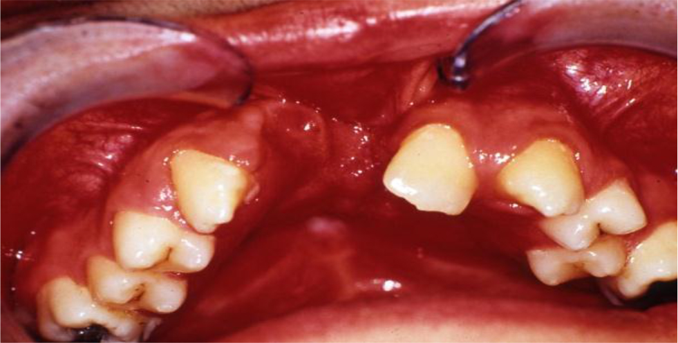
Figure 1. Absence of premaxillary area.
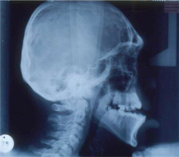
Figure 2. Lateral cephalometric radiography Short Facial Height, concave profile.
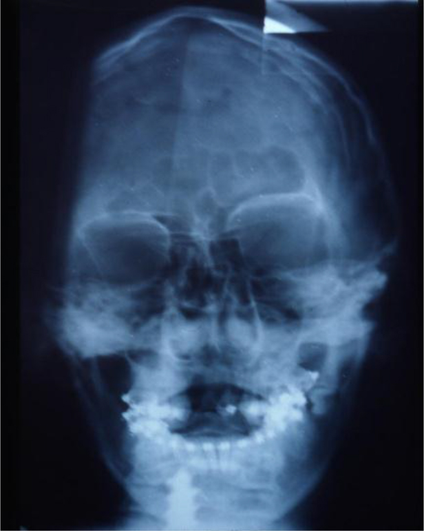
Figure 3. Posterior-front cephalometric radiography. Absence of premaxillary area, infraorbital bony distance.
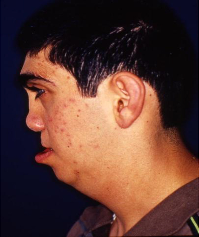
Figure 4. Concave profile.
His karyotype was normal, 46, XY (G-bands).
Panoramic radiograph
Absent teeth
52, 51, 61, 62
18 13, 12, 11, 21, 22, 23 28
48 45, 43, 41 31 33 35 38
Cephalometrics
Patient 10.5 -year-old Father 35-year-old
Cranial base
S-N 71.8 mm (-2.2 SD) 70 mm -3.5 SD
S-Ba 44 mm (-0.6 SD) 48 mm norm
S-N-Ba 129.6 dg (0.1 SD) 126 dg norm
SN-FH 151 dg (3.1 SD)
ANS-PNS 43 mm (-5.4 SD) 50 mm -3.0 SD
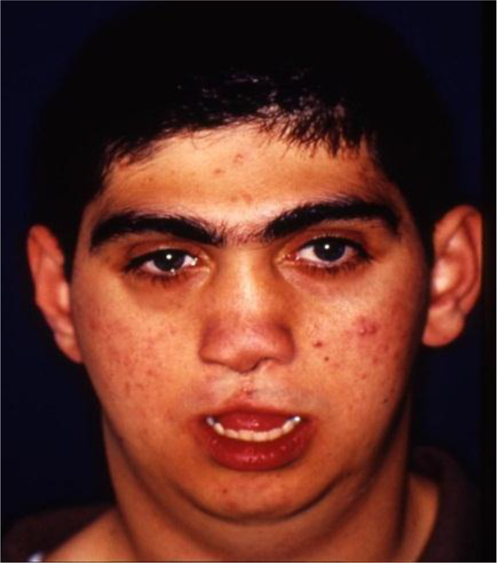
Figure 5. Surgically corrected congenitally absent philtrum.
Skeletal Relations
Facial Angle 90.6 dg (2.3 SD) 88 dg 3.0 SD
(PN-FH)
Lande’s Angle 81.0 dg (-1.2 SD) 91 dg 3.0 SD
(AN-FH)
Convexity -17.8 dg (-5.6 SD)
180-(NAP)
Vertical Analysis
Mandibular Plane 20.2 dg (-1.9) 23 dg
Y-Axis 51.8 dg (-2.5)
UFH (N-ANS) 47.1 mm (-2.0) 61 mm norm
TFH (N-Me) 106.7 mm (-3.0) 132 mm norm
UFH/TFH 44.2% 43.93% 46.21% SNA 82 dg norm
SNB 80 dg norm
ANB 2 dg norm
|
Anterior Cranial Base: |
Severely short |
|
Posterior Cranial Base: |
normal |
|
Saddle Angle: |
normal |
|
Palatal Plane: |
Severely short |
Maxilla: Mildly retruded to forehead severely protruded to forehead well related to anterior cranial base
Mandible: Prognathic to forehead severely protruded to forehead well related to anterior cranial base
Convexity: Severely decreased; concave skeletal profile Overclosure tendency Maxilla and mandible well related to each other Bony interorbital Distance: 18 mm 23 mm
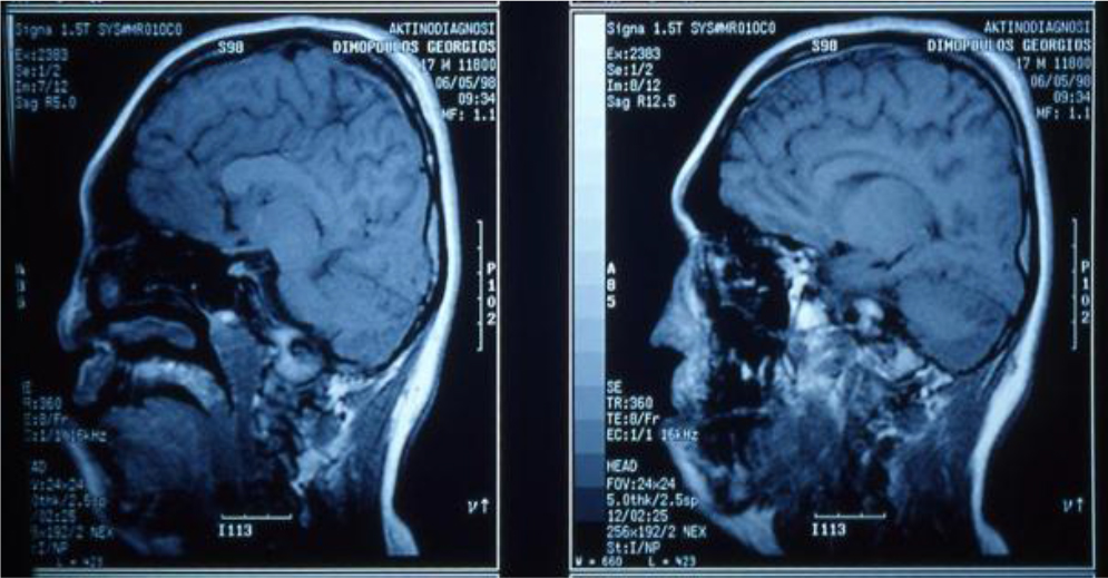
Figure 6. Lateral MRI tomography showing posterior displacement of maxillary sinuses.
Reference
- Gorlin RJ, Cohen Jr, MM Hennekam RCM (2001) Syndromes of the Head and Neck, OXFORD Universal Press. Rieger syndrome (hypodontia and primary mesodermal dysgenesis of the iris). Pp: 1181–1183.