Monthly Archives: February 2020

Measurement and Clinical Significance of Novel Inflammatory Biomarkers in Patients with Psoriasis and Cardiovascular Risk Factors for the Primary Care Practitioner

Summary

Large part of the current research in the field of psoriasis is directed towards identifying and characterising new biomarkers, for both diagnostic and prognostic purposes at clinical settings. This will help in developing prompt and effective therapeutic approaches, particularly to manage the cardiovascular risk factors often associated with psoriasis, to reduce cardiovascular morbidity.

This review highlights the biomarkers that have been identified to date, that are accessible and very useful to primary care physicians (PCPs), because, in most cases, these are the first indicators of psoriasis in the patients. We have focussed on the inflammatory and oxidative stress pathways to identify inflammatory biomarkers that link cardiovascular disease (CVD) risk with psoriasis, in both sexes. In addition, we have separately collated the information specific to women, unlike previous reviews.

Our review also emphasises the importance of staying abreast of the advances in the field, to understand the comorbidities of psoriasis. With new therapeutic approaches that offer novel treatment options, chances of detecting the comorbidities, even in their subclinical phase, improve. This can lead to accelerated clearance of skin lesions, and improved quality of life for the patients.

Key words

Psoriasis comorbidities, cardiovascular risk factors, inflammatory biomarkers, new biological treatments, early clinical diagnosis of psoriasis, primary health care

Introduction

Psoriasis vulgaris is a chronic, immune-mediated, inflammatory, polygenic skin disorder. Its prevalence is approximately 2% in the general population. It has a universal occurrence with a very similar proportion in men and women [1]. It can appear at any age, but two peaks in age of onset have been reported: the first between 20 and 30 years, and the second between 50 and 60 years [2].

Plaque-type psoriasis is the most frequent presentation representing 90% of cases and clinically manifests as well-demarcated erythematous plaques covered by silvery-white scales. They are located mainly on the extensor surfaces of the extremities, scalp, sacral area, and umbilicus. Psoriasis has a notable effect on quality of life; patients often experience depression, anxiety, and stigma as well as suicidal behaviour [3].

As Surcel et al. and Benson et al. [4,5] the knowledge of the pathogenesis of psoriasis has evolved over the years. Currently, the roles played by interleukin (IL) -23, IL-17, IL-22, T helper (Th) -17 cells, Th-22 cells, regulatory T cells, transforming growth factor (TGF) -β1 and IL-10 in the pathogenesis of the disease have been highlighted [4, 5].

It has been reported by Fernández-Armenteros et al. [6] that psoriasis is an independent risk factor for the development of coronary artery disease and acute myocardial infarction. Thus, patients with moderate and/or severe psoriasis have a higher risk of developing ischemic heart disease or cerebrovascular disease than the general population.

Kimball et al. [7] conducted a study with 1,591 patients with moderate or severe psoriasis, and, based on the Framingham table, they estimated the 10-year risk of coronary heart disease and stroke in this population. They verified that for patients with a Psoriasis Area and Severity Index (PASI) score (>20), the risk was estimated at 12.2% and 8.7%, respectively.

Hu et al. [8] analysed the risk of stroke in patients with psoriasis. They found that psoriasis, in mild and severe forms, is an independent factor directly related to the development of stroke. They found no relationship between psoriasis and suffering or depression.

In a thorough study, different comorbidities are listed that may accompany psoriasis, noting that several treatments target interleukin (IL) 17. However, it is still unclear whether they should be used in patients who are at a higher risk of developing CVD. There are some studies that describe its efficacy in psoriasis patients who are affected by myocardial infarction, hypertension, and chronic renal failure [9].

Anti-IL17A/F treatment has been tested in a randomized, double-blind, placebo-controlled Phase 1 clinical trial. Changes were observed in liver enzyme levels, and clinical readouts, such as vital signs, electrocardiograms, or body weight. In this trial, no deaths were reported [10].

“Author last name” briefly describes biomarkers for systemic inflammation, noting that some are prognostic biomarkers, and that some are diagnostic biomarkers. A complete review of biomarkers is described here in our manuscript.

Thus, psoriasis can be considered as the amplifier of the inflammatory march if the metabolic disorder appears before the diagnosis of psoriasis [11].

Patients with psoriasis have been found to have increased arterial stiffness. Increased risk of hypercholesterolemia, atherosclerosis, metabolic syndrome, and insulin resistance may also develop after chronic vascular inflammation; improvement of psoriasis can lead to a reduction in aortic vascular inflammation [12]. Additionally, patients with psoriasis are more likely to have subclinical atherosclerosis, greater carotid artery intimal thickening, and substantial endothelial dysfunction than the general population. The inflammatory nature of atherosclerosis and psoriasis has been highlighted recently; both are associated with T lymphocyte-mediated adaptive immune events and mechanisms involving innate immunity [13].

Alexandroff et al. [14] reported that obesity and psoriasis produce subclinical inflammation characterized by an increase in the levels of pro-inflammatory cytokines such as IL-6, TNF-α, and C-reactive protein (CRP).

Schäkel et al. [15] focused on the innate and adaptive immune system and found interactions among neutrophils, macrophages, TH1 lymphocytes, monocytes, platelets, and IL-17a, which have been implicated in the pathophysiology of psoriasis and atherosclerosis.

Chronic inflammatory systemic diseases (CISDs) are part of a spectrum of chronic diseases in which inflammation plays an important role in pathogenesis. CISDs and cardiovascular diseases, including atherosclerosis, share common pathogenic features in which inflammatory cytokines play an important role. Common pathogenic features include immunological processes (diseases mediated by Th-1 cells), inflammatory cytokine profiles, and the presence of inflammatory markers both locally and systemically. Activation of these inflammatory cells, together with the release of inflammatory cytokines and proteases, contributes to the development of psoriatic lesions and plays a major role in the breakdown of atherosclerotic plaques [14].

Boehncke et al. [16] found that reduced levels of adiponectin are associated with insulin resistance and several adverse vascular events and are inversely correlated with serum CRP in obese patients with diabetes and those with coronary artery disease. Risk factors for cardiovascular disease (obesity, insulin resistance, and dyslipidaemia) are associated with decreased levels of adiponectin and high levels of leptin. Elevated levels of leptin in patients with psoriasis are associated with overweight and obesity, but not with the severity of psoriasis [17].

The assessment of cardiovascular risk through scales helps in therapeutic decision making and in cardiovascular prevention. The following necessary variables were collected for the calculation of coronary risk using both the Registre Gironí del cor (REGICOR) and Systematic Coronary Risk Evaluation (SCORE) scales: age, sex, total cholesterol, high density lipoprotein (HDL) cholesterol, systolic blood pressure, diastolic blood pressure, smoking history, and antecedent of diabetes mellitus (DM).The calculation of the risk of cardiovascular death by SCORE is based on the figures of total cholesterol and for countries of low risk, it was based on the following: age, sex, total cholesterol, systolic blood pressure, and smoking history [18] or the JUPITER study where HDL was an inverse predictor of incident events and biomarker of residual risk [19].

However, there is a discrepancy in the estimation of these scales for cardiovascular risk since they have limitations. Comparative studies were performed among the SCORE, REGICOR, and Framingham scales in our setting, and discrepancies were found in the detection of high risk and therapeutic indications (SCORE favours intervention in women with hypertension and Framingham in men with dyslipidaemia). The SCORE has the aggravating circumstance that it only estimates cardiovascular mortality and not morbidity [18–20].

Other scales used to determine cardiovascular risk were the American Framingham and POOLED cohort [21]. In patients with moderate-severe psoriasis, it is probably convenient to multiply the score obtained according to Framingham or SCORE by 1.5 [22].

In Spain, the only valid scale is REGICOR, but it only measures coronary risk. European countries support the SCORE scale but it only measures mortality and does not include people over 65 years of age. Currently, some authors consider the English scale promoted by the National Institute for Health and Care Excellence (NICE) as one of the most complete. The scale is called QRISK. It is available online at https: //qrisk.org/three [23] is the 3rd version [24].

In any case it is convenient follow the guidelines proposed by Dauden et al. [2]. Following the Spanish recommendations, screening for cardiovascular risk factors should be performed every 6 months in patients with systemic treatment and yearly in patients with topical treatment.

In the study conducted by Cea-Calvo et al. [26], the presence of undiagnosed cardiovascular risk factors (CVRF) in patients with psoriasis was investigated. For this, 368 patients were recruited, with an average age of 48 years, 96% with psoriatic plaque, 23% with psoriatic arthritis, and 4% with other forms of psoriasis.

Through the measurement of blood pressure and fasting blood test, new cases of cardiovascular risk factors (CRFV) were detected in more than 27% of patients. In addition, the 10-year cardiovascular risk was similar to that of patients with known CVRF [26] macrophages.

The PCPs are important in the evaluation of cardiovascular risks in patients with psoriasis, because often they are the only physicians to treat and follow up with these patients. Their role can be more effective, if they possess sound knowledge of these pathologies and training to handle and treat them [27]. Research on this disease is primarily directed towards identification and characterization of new biomarkers. In this review, information about the main diagnostic and prognostic biomarkers, and their responses to different treatments, were collected from the patients of psoriasis with cardiovascular risk. These data can be useful in clinical practice.

Correlation between Psoriasis and Cardiovascular Functional Indices

Moore et al. [28] reported that the development of an atherosclerotic plaque is one of the most important risk factors. It is a multistep process and inflammation contributes to both the development of atheroma itself and the eventual rupture of the plaque; the first step is altered endothelial function with a change in the normal homeostatic responses; the next step is the recruitment of macrophages into the developing lesion and the accumulation of lipid-laden macrophages (foam cells) to form a fatty streak and the formation of a more advanced fibrous lesion, which is covered by a fibrous cap. Then, it gradually develops into an advanced and complex lesion, continuing in thinning of the fibrous cap to create a potentially unstable plaque that can eventually rupture, which leads to thrombosis and clinical evidence of vascular occlusion [28].

Family history of atherosclerosis, metabolic syndrome, diabetes, obesity, increased levels of inflammatory molecules (Figure 1) [29], discontinuity in the follow-up of patients [30] with newly diagnosed hypertension, diabetes, and hypercholesterolemia were associated with higher cardiovascular events, and associated mortality, escalating the financial cost of the health care.
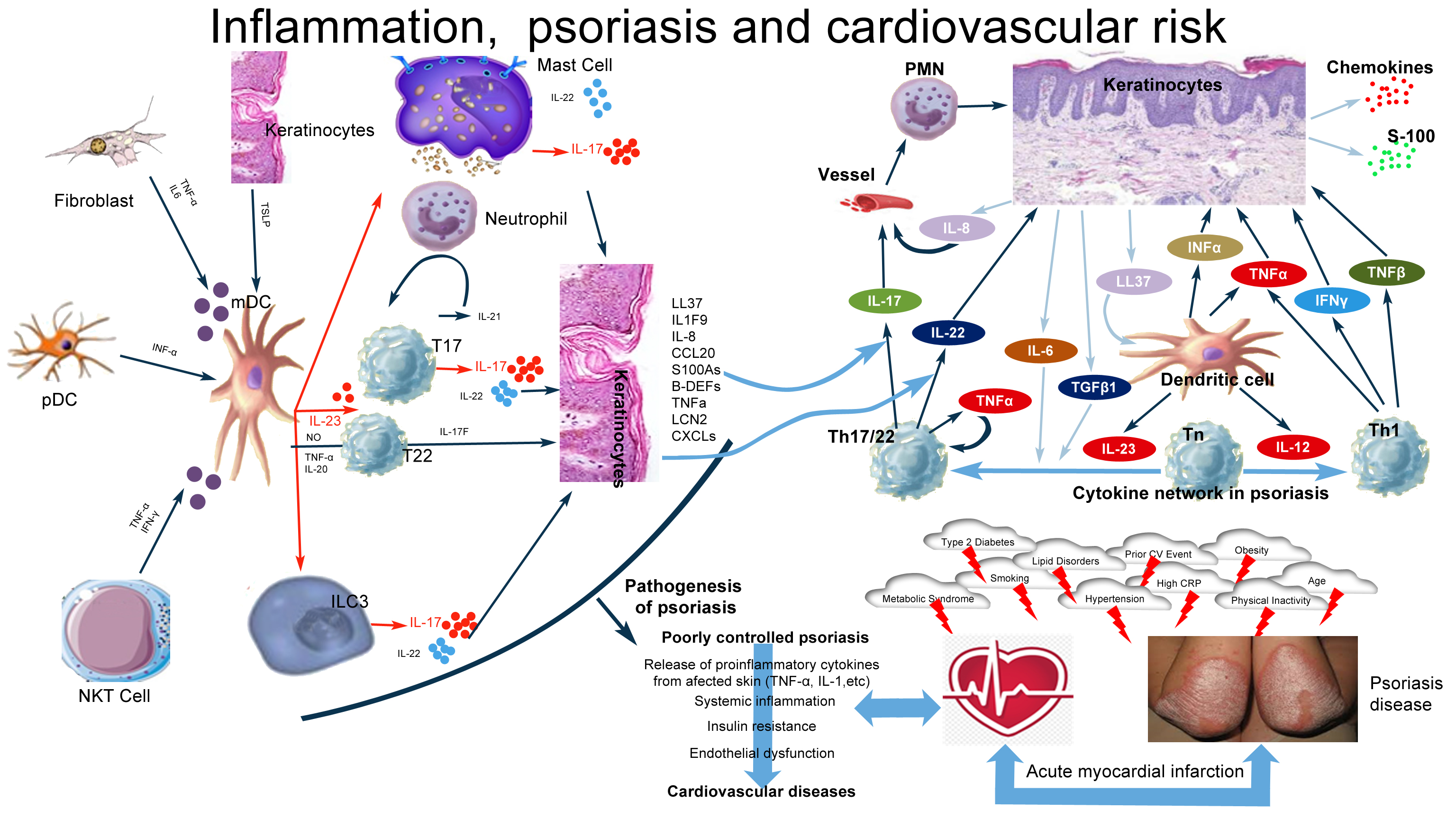
JCRM Carmen Rodriguez-Cerdeira - 2020-302_F1

Figure 1.

Histopathological changes observed in psoriasis are due to important and complicated interactions between keratinocytes, dendritic cells, T lymphocytes, neutrophils, and mast cells. IL-23, IL-17, IL-22, Th-17 cells, Th-22 cells, regulatory T cells, TGF-β1, and IL-10 are involved in the pathogenesis of the disease. The IL-23/IL-17 axis is the main immune pathway in the pathogenesis of psoriasis. Multiple factors induce mDC activation with consequent IL-23 production (IFN-α, TSLP), which, in turn, mainly stimulates T-cell subsets and also ILC3, mast cells, and neutrophils, which secrete IL-17. Other cytokines derived from T cells, mast cells, and ILC3 (IL-22, IL-17F, and IL-21) and from mDCs (TNFα, NO, and IL-20) are responsible for the development of psoriasis. T17 and T22 cells represent all T-cell subsets producing mainly IL-17 and IL-22.

Chronically released proinflammatory cytokines in patients with poorly controlled psoriasis pass into the circulatory system and potentiate and perpetuate systemic inflammation. This systemic inflammation causes obesity, hypertension, dyslipidemia, and type 2 DM . Additionally, this leads to an increase in insulin resistance, endothelial dysfunction, and the onset of cardiovascular diseases.

 Environmental factors studied by Balak et al. [31] included infectious agents, a high-fat diet, smoking, alcohol consumption, low levels of antioxidants, and lack of exercise, as well as psychological stress, certain medications (antimalarial drugs, beta-blockers, lithium, and nonsteroidal anti-inflammatory drugs), and a history of skin disorders (e.g. psoriasis).

However, there is little data about subclinical alteration of myocardial function and adjacent vascular changes in patients with mild psoriasis. Milaniuk et al. [31] used two-dimensional stress echocardiography (2D-SE) for the quantification of alterations in all dimensions of the left ventricle. They reported that an increase in stiffness negatively affects cardiac function.

Dattilo et al. [33] evaluated cardiac performance and vascular rigidity in patients who had mild psoriasis and had no other risk factors or were undergoing treatment. The patients came from the department of Dermatology where they went periodically for follow up. Finally, 33 patients were included in the study sample. The control group consisted of healthy volunteers. The researchers found a significant correlation between global longitudinal strain and patient age. They also found that arterial stiffness values were significantly higher in psoriatic patients [33].

A study conducted by Sutton-Tyrrell et al. [34] showed arterial stiffness, a characteristic finding in patients with CISDs, is one of the most important determinants of increasing systolic blood pressure and pulse pressure; thus, it is a major cause of cardiovascular complications and events.

Ardic et al. [35] reported comparable left ventricle dimensions, wall thickness, and ejection fraction between patients with psoriasis and healthy controls.

Goga et al. [36] determined the presence of preclinical cardiac dysfunction in a cohort of 52 psoriatic patients without cardiovascular risk factors. They compared them with a control group. Both groups underwent echocardiographic conventional and tissue Doppler (TDI) studies. Left ventricular diastolic dysfunction was found in 36.5% patients in the psoriasis group versus 0% in control group. In addition, it was found that patients with psoriasis have a significant increase in mitral regurgitation [36].

Balbul-Sen et al. [37] conducted a cross-sectional and observational study with 65 patients with psoriasis and a control group. They verified that carotid intima-media thickness (CIMT) is a potential indicator of subclinical atherosclerosis in patients with psoriasis. Epicardial fat thickness (EFT) is proposed as a new cardiometabolic risk factor. They stated that EFT and CIMT are increased in patients with psoriasis, and that echocardiographic EFT will be correlated with CIMT in patients with psoriasis. This shows that echocardiographic study through EFT may be a marker of subclinical atherosclerosis and increased cardiovascular risk in patients with psoriasis.

Another comorbidity of psoriasis considered by Gisondi et al. [38] is the non-alcoholic fatty liver, which is currently considered to be the hepatic manifestation of metabolic syndrome. This condition is linked to an increased risk of future cardiovascular events independent of conventional risk factors [39].

Schiffrin et al. [40] found that a decreased flow velocity in the brachial artery indicated a risk of developing coronary artery disease.

El-Mongy et al. [41] reported that patients with psoriasis had more prevalent valvular regurgitation, abnormal diastolic relaxation, left ventricular hypertrophy, left ventricular diastolic dysfunction, left ventricular wall motion abnormalities. Mitral valve and tricuspid valve prolapse were found to be more prevalent in patients with psoriasis in a few studies, but in other studies, these results were not confirmed.

Echocardiographic findings from Biyik et al. [42] demonstrated that compared with the control group, patients with psoriasis had greater aortal diameter, greater septal and posterior wall thickness, more frequent tricuspid and mitral regurgitation, and increased systolic pressure in the right ventricle. Early atherosclerotic echocardiography predictors were found in the patients with psoriasis.

Goyal et al. [43] reported that psychological stress and depression have been increasingly in recent years and have been associated with multiple traditional cardiovascular risk factors. The amygdala regulates physiological and behavioural changes in response to stress and fear; using 18-fluorodeoxyglucose positron emission tomography computed tomographic (FDG PET/CT) imaging, the resting metabolic activity in the amygdala can be reproducibly assessed. Measures of stress-related neural activity are associated with anxious temperament and are upregulated in anxiety disorders, chronic stress, and depression. In 2017, Tawakol et al. [44] studied the importance of amygdalar activity (AmygA) with increased bone-marrow activity and arterial inflammation but they studied patients who did not have psoriasis. Goyal concluded that AmygA is strongly associated with aortic vascular inflammation (VI) and risk of major cardiovascular events. This association is partially mediated by upregulated hematopoietic system activity (HMPA). Psoriasis patients have elevated rates of anxiety and depression, and is also an independent factor directly related to development of CVD.

A retrospective study was conducted by Shiba et al. [45] to analyse the relationship between psoriasis and myocardial infarction, diabetes, and dyslipidaemia. They included more than 100,000 patients of all ages and performed multiple statistical analysis with the following results: patients with psoriasis have a higher prevalence of hypertension, DM, myocardial infarction, and hyperlipidaemia. There is an independent association between psoriasis and myocardial infarction.

Correlation between Cardiovascular Risk and Psoriasis Severity Indices

The importance of the severity of psoriasis in the development of cardiovascular disease is also supported by evidence of a correlation between PASI values and insulin secretion. Insulin resistance is a characteristic feature of metabolic syndrome. In patients with psoriasis, a statistically significant correlation has been observed between serum levels of resistin (a cytokine that is elevated in insulin resistance situations) and PASI [46]. These data indicate that the severity of psoriasis is related to an increased risk of cardiovascular complications and point to the pathogenic importance of a state of chronic inflammation in the development of vascular disease in these patients.

Prodanovich et al. [47] found a relationship between the severity of psoriasis with the risk for atrial fibrillation and ischemic cerebrovascular disease, with an atrial fibrillation incidence of 3.03 in patients without psoriasis compared to 5.96 in patients with severe psoriasis; similar figures were detected for ischemic cerebrovascular disease.

The risk score measured with the Framingham scale by Eder et al. [47] was significantly higher in patients with chronic plaque psoriasis than in the control group (grouped by age and sex). In patients older than 50 years, there was no correlation between the risk score and the severity of PASI [48].

To relate the severity of psoriasis to vascular inflammation, Naik et al. [49] recruited 60 patients with psoriasis and 20 controls, and measured the severity of the disease through PASI and vascular inflammation with the use of FDG PET/. They demonstrated that severe psoriasis is associated with atherosclerotic disease, and the presence of the disease alone increases cardiovascular risk, coupled with concomitant risk factors [49]. The contribution of PASI score in predicting vascular inflammation (aortic target-to-background ratio) beyond Framingham risk score and high-sensitivity C-reactive protein (hsCRP) was determined using likelihood ratio testing in nested models PASI score. The authors found that the severity of psoriatic skin disease increases vascular inflammation independently [49].

In a review by Prey et al. [50], they investigated the psoriasis cases (with different degrees of severity) and cardiovascular risk factors described in the literature between 1980 and 2009. Finally, 18 articles were selected from the main medical databases. There was an association between diabetes and psoriasis in 11 studies. The risk of diabetes was higher in patients with a moderate or severe degree of psoriasis [51, 52]. Three studies showed an association between hypertriglyceridemia and psoriasis, and only one study showed an association between psoriasis and total cholesterol levels. None of the studies showed an association between psoriasis and risk of dyslipidaemia or HDL cholesterol levels.

The risk of hypertension was described in 10 studies compiled by Herron et al. [53] but a relationship between the level of risk and the degree of severity of psoriasis was not established. Eight articles showed the risk of obesity in psoriasis and reported that the risk increases directly depending on the severity of psoriasis. All articles showed an increased risk of metabolic syndrome (MS) in patients with psoriasis [53].

Chronic stress-related neural activity as measured by AmygA was higher in patients with severe PASI. Increased AmygA was related to the severity of psoriatic skin disease and subclinical cardiovascular disease (CVD). The amelioration of psoriasis with therapy was accompanied by reductions in AmygA, HMPA, aortic vascular inflammation, and stabilization of non-calcified coronary plaque burden (NCB). Stress affects the endocrine system, leading to upregulation of circulating catecholamines, glucocorticoids, and inflammatory cytokines, which subsequently leads to CVD. Psychological stress should be considered in evaluating CVD risk, especially in inflammatory disease states. The effect of reducing inflammation, as proven by improvement in PASI severity, on AmygA, HMPA, aortic VI, and NCB, [43].

In a study conducted by Uyar et al. [54], the mean serum concentrations of fetuin-A, alanine aminotransferase (ALT), and rheumatoid factor levels were found to be higher in patients with psoriasis. Median CIMT levels were significantly higher in patients with mild-to-moderate psoriasis compared to controls, but no correlations were found among the serum fetuin-A levels, CIMT levels, and PASI scores.

Rosa et al. [55] showed that 75% of patients with moderate or high risk presented a with a PASI below 10. Thus, although psoriasis patients had an increased cardiovascular risk, there was no relationship between severity of the cutaneous manifestations and a higher risk.

In a study by Gisondi et al. [56], using the Framingham risk score, they found the severity of cutaneous manifestations, measured by PASI, was associated with an increased cardiovascular risk.

Sorokin et al. [57] demonstrated that patients with psoriasis have a relative and absolute increase in circulating monocyte aggregates as well as an increase in intermediate monocytes, which correlates with an increase in the severity of PASI.

Sphingolipids, specifically ceramides (CER) and sphingosine-1-phosphate (S1P), are signal molecules that are responsible for regulating cell growth, apoptosis, and immune reactions. Myśliwiec et al. [58] demonstrated that circulating levels of CER and S1P in plaque-type psoriasis were associated with disease activity through the PASI measure.

Women, psoriasis and CV risk

Interestingly, there were few instances of psoriatic women with increased risk of developing CVD. Some studies grouped patients based on sex, however, CVD risk was not always reported with patient sex. In this section, we collected information from the few studies that observed CVD risk in psoriatic women.

In Denmark, Blegvad et al. [59] studied 2,435 women with psoriasis (2.90%) and 81,388 women without psoriasis and concluded that psoriasis is significantly associated with hypercholesterolemia and hypertension. Women with psoriasis have an increased risk of developing cardiometabolic disorders in early adult life. Screening for classic risk factors such as hypertension, hypercholesterolaemia, and hyperglycaemia, is advised. Additionally, preventing type 2 diabetes may be important when treating patients with psoriasis, particularly in younger patients.

In a study by Pietrzak et al. [60], 432 patients with psoriasis and a control group of 546 were selected. Both psoriatic and control patients showed that CVD occurrence increased with age and concluded that: a) the incidence of psoriasis and CVD in women increases with age, b) it is unclear whether menopause increases the risk of psoriasis, c) it is unclear whether menopause and resulting hormonal disturbances are independent CVD risk factors, and d) investigating whether hormone replacement therapy reduces CVD risk in psoriatic patients may be important.

Li et al. [61] conducted several studies on psoriatic women and the risk of developing CVD. In particular, the study published in Nurses’ Health Studies (NHS) collected epidemiological information about the health status of women in different diseases, showing a strong relationship between psoriasis and CVD. In another study, there was a significantly higher risk of type 2 diabetes in psoriatic patients, and that this occurred particularly among younger women who had developed psoriasis at an early age [62]. Some of these patients eventually developed CVD. Li et al. [63] also showed that women with psoriasis had increased non-fatal CVD resistance, especially myocardial infarction. Therefore, women who developed psoriasis earlier, or those who have had it for a long time, presented an increased risk of myocardial infarction.

Prognostic Biomarker

The onset of psoriasis at an early age increases the risk of myocardial infarction. This appears to be due to an increase in the time of exposure to a chronic systemic inflammation. In a cross-sectional study with 301 participants, Kumdai et al. [64] found that certain HLA antigens, including A30, B50, Cw6, and DR7.20, were associated with early-onset psoriasis.

On the other hand, Wongpiyabovorn et al. [65] reported that IL-10 distal promoter polymorphisms are usually associated with late-onset disease.

Oxidized LDL (OxLDL) is very important in the exacerbation of atherosclerosis; thus, it is an important factor in the treatment of cardiovascular disease. In psoriasis, the efflux capacity of cholesterol is decreased, which changes after treatment. Activated oxidation lipid mediators (OMLs) abound in psoriatic skin, playing a critical role in the process of initial inflammation and resolution. A study by Sorokin et al. [57] with 252 patients with psoriasis and a control group measured LDL with modified oxidation, HDL, lipoprotein A, cholesterol efflux capacity, and size and number of lipoproteins. They observed an increase in oxidized Lp (a), Lp (a), and oxidized HDL. The activity of paraoxonase-1 was increased by possible compensatory oxidative effect. It was concluded that psoriasis is associated with increased levels of some OMLs as opposed to healthy individuals. OxLDLs have a direct association with the non-calcified plaque. The authors suggest that serum levels of OML could be useful as early markers in atherosclerosis by coronary computed angiography, which allows the quantification and characterisation of non-calcified plaque in the coronary arteries [57].

Dinić et al. [66] recruited 66 patients with chronic psoriasis plaque. The control group consisted of 20 patients with generalized eczema and 20 healthy controls, all of whom were between 18 and 60 years old, had no associated comorbidities, and underwent systemic treatment. Blood samples were taken and analysed. The values for the following were obtained: erythrocyte sedimentation rate (ESR), fibrinogen, PCR, Dimer-D, serum amyloid A, Apolipoprotein (Apo) A1, Apo B, Apo B / Apo A1 ratio, serum concentration of uric acid (SUAC), fasting insulin, Peptide-C, creatinine clearance obtained by Cockroft-Gault formula, microalbuminuria and proteinuria in 24 h. Colour duplex Doppler ultrasound was also performed to measure the following: arteriovenous resistance; the thickness of the medial intima of the common carotid artery distal to the posterior wall of the carotid bifurcation; the femoral artery and its resistance and pulsatility indices; and maximum flow velocity of the femoral and brachial arteries. B-mode ultrasound was used. Patients with psoriasis presented with the parameters associated with cardiovascular risk, in addition to metabolic syndrome, even in the absence of other risk factors [66].

Three distinct monocyte populations have been identified and genotyped within human blood: classical monocytes (CD14 ++ CD162), intermediate monocytes (CD14 ++ CD16 +), and nonclassical monocytes (CD14 + CD16 ++). Among the results obtained by Golden et al. [57], it was found that patients with psoriasis have a higher percentage of intermediate circulating CD14 + CD16 + monocytes compared to controls. This was obtained through peripheral blood samples, and/or biopsies in healthy controls and in patients with psoriasis. Systemic or topical therapy were not used for the preparation of mononuclear cell preparations in peripheral blood. It was also shown that the classical monocytes subset (CD14 ++ CD162) correlates negatively with PASI, and the nonclassical monocytes (CD14 + CD16 ++) do not show any correlation [67].

The vascular uptake of fluorodeoxyglucose (FDG) is associated with unstable atherosclerotic plaques. A relationship was found between arterial inflammation detected by FDG PET/CT and the presence of high-risk coronary plaque, which is mainly composed of cells of the psoriatic plaques. This suggests a potentially shared immune mechanism and demonstrates the role of neutrophils in the perpetuation of psoriasis and their comorbidities; the downregulation of CD16 and CD62L in this sample indicates a decreased ability to mediate inflammatory damage and a greater activation state. It was also reported that S100A8/A9 proteins may serve as mediators or by-products in one of the potential pathays linking psoriasis severity and arterial inflammation, confirming the role of neutrophils in cardiovascular disease associated with psoriasis [49].

Ryan at al. [68] found microparticles present in the atherosclerotic plaque breakdown, which causes cardiovascular events. They also showed an increased concentration in these microparticles in patients with psoriasis.

Homocysteine is an amino acid produced in the liver that is involved in the pathogenesis of several diseases, including psoriasis. It increases the levels of oxidative stress and inflammation, and induces apoptosis of endothelial cells, which favours atherosclerosis; atherosclerosis is the most common cause of cardiovascular pathology such as myocardial infarction or heart failure. Homocysteine levels can be used as predictors for cardiovascular risk, and they are directly related to the severity of psoriasis [69]. Homocysteine plays an important role in the DNA methylation cycle, which is a process related to epigenetic mechanisms. Wang et al. [70] reported that several studies have observed that hyperhomocysteinemia reduces levels of DNA methylation, which is related to the pathogenesis of psoriasis. Genes that encode proteins such as HLA-DRB1, protein tyrosine phosphatase 1 (SHP-1), or different high proliferative potential colony-forming cells (HPP-CFCs) are hypomethylated.

Qi at al. [71] reported that methylene tetrahydrofolate reductase (MTHFR) participates in the irreversible conversion of a cofactor that participates in the remethylation of homocysteine to methionine. Only one polymorphism of the MTHFR gene (677C> T) is related to psoriasis, and there are reports of the relationship between this polymorphism and cardiovascular diseases. This polymorphism is not a risk factor in the pathogenesis of psoriasis, but it can influence the severity of psoriasis.

Psoriasis in combination with obesity/MS produces greater systemic inflammation and greater oxidative stress, which promotes endothelial dysfunction and the formation of atherosclerotic plaques, which increase the risk of cardiovascular disease. Kaur et al. [72] found that obesity and psoriasis produce subclinical inflammation characterized by an increase in the levels of pro-inflammatory cytokines such as IL-6, TNF-α, and CRP. The levels of CRP and IL-6 are related to the severity of psoriasis.

Psoriatic skin disease severity showed a linear relationship with AmygA, and was associated positively with subclinical cardiovascular disease, a higher Framingham risk score, more insulin resistance, and higher CRP levels. When psoriatic skin disease severity improved, there was a significantly concurrent decrease in AmygA, bone marrow activity, and aortic elasticity, and found beneficial effects of skin disease clearance on the amygdala and aorta after one year of treatment [73, 74].

The CRP is an acute phase reactant protein produced from hepatocytes within hours after being stimulated from infection, inflammation, or tissue damage. Its levels decrease rapidly after the stimulating factor has been eliminated or has responded to treatment. Uaratanawong et al. [75] showed that the hs-CRP level was significantly higher in the psoriasis group with a significant correlation between hs-CRP and PASI score after controlling for other confounding factors. A hs-CRP level greater than 0.69 mg/L could be the marker of worsening psoriasis. The keratinocytes in psoriasis lesions secrete tumour necrosis factor-α and interleukin-1, which can stimulate hepatocytes to produce hs-CRP. Furthermore, hs-CRP level can predict cardiovascular risk, including cerebrovascular and peripheral arterial disease (hs-CRP less than 1 mg/L as a low risk, 1–3 mg/L as intermediate risk, and more than 3 mg/L as high risk for cardiovascular event). Although hs-CRP is not a specific marker and additional work will be imperative to validate this marker, it shows the inflammatory burden of psoriasis and could be a potential marker that can be used for grading psoriasis severity with great cost-effectiveness.

When assessing coronary artery disease and generalized atherosclerosis, CIMT is a good indicator of an increased risk of subclinical atherosclerosis. Fetuin-A is a serum glycoprotein that is synthesized mainly by hepatocytes, increases insulin resistance and dyslipidaemia and reduces ectopic calcification in vascular disease. There are reports on fetuin-A interacting with transforming growth factor (TGF)-β and epidermal growth factor (EGF), which play an important role in the pathogenesis of psoriasis [76].

Ix et al. [77] reported that lower fetuin-A levels initiate calcium deposition within the vasculature but not atherosclerosis directly, and that they are independently associated with greater coronary artery calcification severity but not peripheral arterial disease. They observed a correlation between serum fetuin-A levels and fasting glucose, as well as between fetuin-A and LDL cholesterol, so it is recommended that physicians should measure CIMT rather than the less appropriate serum fetuin-A values when assessing the cardiovascular disease risk of patients with psoriasis.

Serum adipokines have been considered in the pathogenesis of the psoriasis and have been demonstrated to be associated with insulin resistance. Adiponectin, resistin, and leptin have possible antiatherogenic and anti-inflammatory properties by regulating energy homeostasis and increasing insulin secretion and sensitivity. Hyperleptinemia has contributed to cardiovascular risk in patients with the metabolic syndrome; adiponectin and leptin are predictors of microvascular complications in patients with type 1DM [17].

A study by Yazıcı et al. [78] included 65 patients with type 1 DM. Leptin levels were reported to be decreased in adult patients with type 1 DM without association with CIMT. Resistin levels were decreased in adult patients with type 1 DM and increased in patients with type 2 DM with CAD and was correlated positively with CIMT; increased resistin levels in patients with type 1 DM seem to be suggestive of subclinical atherosclerosis. Leptin was associated with CIMT in patients with type 2 DM. Thus, after adjusting for other known predictors, only resistin was associated with subclinical atherosclerosis in this group of patients. Ataseven et al. [79] suggest that vaspin and VAP-1 may play a role in the pathogenesis of psoriasis and can be used as markers of the disease. Therefore, vaspin levels can be used as a marker of the severity of the disease.

Diagnostic Biomarkers

A strong expression of chemerin was observed by Nakajima et al. [79] in pre-psoriatic skin adjacent to active lesions and early lesions in the dermis. They also observed the presence of CD15+ neutrophils and CD123+/BDCA-2+/ChemR23+ pDC. In skin from chronic plaques, chemerin expression was lower. Additionally, the segregation of neutrophils in epidermal microabscesses and a few plasmatoid dendritic cells (pDCs) in the dermis were observed [80].

Chemerin stimulates the chemotaxis of pDCs and neutrophils, and high systemic chemerin level was found as an independent marker of the metabolic syndrome. Recently, Albane   si et al. [81] reported a strong expression of chemerin together with increased numbers of pDCs and neutrophils in the dermis of early psoriasis lesions.

Furue et al. [82] reported that condition which have been proposed include shared genetic factors, common inflammatory pathways, secretion of adipokines, insulin resistance, altered lipoprotein composition and function, angiogenesis, oxidative stress, microparticles, and hypercoagulability; CRP, Th1, and Th17 lymphocytes, resistin and leptin has been found to be elevated, with decreased serum levels of adiponectin. Psoriasis may induce systemic inflammation leading to insulin resistance, endothelial dysfunction, and the development of atherosclerosis and cardiovascular comorbidities in the psoriatic march.

Identification of serum biomarkers (CRP, soluble CD40 ligand, human matrix Gla protein and fetuin-A, serum YKL-40, GlycA, and complement C3) of systemic inflammation is important for assessing cardiovascular risk; adequate treatment for psoriasis may decrease the risk of cardiovascular comorbidities as a result of suppression of systemic inflammation [12].

A group of 74 patients with psoriasis and 65 healthy controls were studied by Vachatova et al. [83]. Individuals with insulin resistance, diabetes, and cardiovascular disease were included in the psoriasis group. None of them had treatments that could influence inflammatory reaction. Patients with psoriatic arthritis were excluded. The patients with psoriasis presented with higher diastolic blood pressure, MS, BMI, and hip circumference compared to the controls. Patients with psoriasis had higher levels of leptin, resistin, and lipoprotein-associated phospholipase A2 (Lp-PLA2) than controls. Among individuals with MS, those in the psoriatic patients group had higher CRP levels of leptin, resistin, and Lp-PLA2 than those of CG. Patients with psoriasis and MS had higher levels of leptin and Lp-PLA2 and lower levels of adiponectin than patients with psoriasis without MS.

The combination of psoriasis and MS increases the expression of inflammatory and anti-inflammatory cytokines and increases the risk of cardiovascular disease. Likewise, adipokines (leptin, adiponectin, and resistin) are involved in the pathogenesis of psoriasis and MS, and the levels of Lp-PLA2 indicate the presence of subclinical atherosclerosis and cardiovascular risk in patients with psoriasis.

Kiluk et al. [84] found that Lp-PLA2 is associated with an increased incidence of cardiovascular events but only a few studies have associated it with psoriasis.

Metabolomics may potentially explain the mechanisms of various diseases, facilitate early diagnosis, and identify possible therapeutic areas.

Wang et al. [85] presented a utility (mmPredict) to determine metabolomic biomarkers, which is complemented by current studies of metabolomic profiles that are based on patient data. This utility reveals the metabolites of psoriasis based on both genetic and phenotypic relevance (genome-phenome). It was shown in the study that mmPredict is more effective in identifying clinically relevant biomarkers in psoriasis than the genome-based approach. They are improving mmPredict to incorporate other types of data such as the phenotype of drugs and high-level diseases observed in humans.

Another study, conducted by Coimbra et al. [86], showed that inflammatory cytokines were involved in the pathogenesis of psoriasis such as IL-22, IL-17, IL-23, IL- 8, TNF-α, and endothelial growth factor.

Ellulu et al. and Rodríquez-Cerdeira et al. [87, 88] reported that reduced levels of adiponectin are associated with insulin resistance and several adverse vascular events and are inversely correlated with serum CRP in obese patients with diabetes and those with coronary artery disease. Risk factors for cardiovascular disease (obesity, insulin resistance, and dyslipidaemia) are associated with decreased levels of adiponectin and high levels of leptin. Elevated levels of leptin in patients with psoriasis are associated with overweight and obesity, but not with the severity of psoriasis.

Excess fatty tissue, due to obesity associated with psoriasis, contributes to atherogenic dyslipidaemia, and several studies have associated atherogenic dyslipidaemia with increased blood levels of total cholesterol, triglycerides, LDL, and apolipoprotein A, and low levels of HDL and apolipoprotein B in patients with psoriasis.

In a study by Asha et al. [89], ox LDL is considered a marker of hyperlipidaemia and atherosclerosis. A few studies have shown increased levels of oxLDL in patients with psoriasis, which was consistent with the BMI. oxLDL/LDL ratio a better predictor for atherosclerotic risk as compared to LDL

Advanced oxidation protein products (AOPPs) and adipokines are also involved in the pathogenesis of endothelial dysfunction and atherosclerosis.

In other study conducted by Haberka et al. [90], patients with non-severe psoriasis had significantly increased impaired endothelial function and CIMT, and significantly increased serum levels of AOPPs and visfatin. Increased oxidative stress and serum visfatin levels are associated with a chronic inflammatory state; increased AOPP levels are associated with endothelial dysfunction (FMD), vascular remodelling CIMT, and increased serum visfatin levels, suggesting that increased oxidative stress is the main pathomechanism involved in cardiometabolic complications found in non-severe psoriasis.

In patients with psoriasis with a risk allele of FTO gene rs9939609, Tupikowska-Marzec et al. [91] suggested that they have an increased risk for more severe psoriasis and obesity, and may develop obesity-induced insulin resistance and therefore, CV risk.

As Barrera et al. [92] Vit D ot only to reduce its clinical severity, but also for delineating the risk profile for co-morbidities cardiac risk factors that may result from psoriasis [93].

Recently, a study in women conducted by Hruska et al. [94] showed a strong relationship between single nucleotide polymorphism (SNP) rs4597342 within the3’untranslated region (3’UTR) of integrin alpha M (ITGAM) with psoriasis susceptibility and severity. The associated risk was observed specifically for allele T of rs4597342, which introduces a binding site for miR-21 that is upregulated during psoriasis [94]. These markers are also linked to IgA nephropathy [95] and type 1 diabetes [96], which could be interesting diagnostic markers for CVD

Treatment Biomarkers

Reviewing the literature, we observed that patients with psoriasis receive suboptimal and disconnected attention and it leads to a decrease in the quality of life. As proposed by Gulliver et al. [97], the concept of treat-to-target, which has already been adopted for other conditions such as diabetes and dyslipidaemia, would be very beneficial for patients with psoriasis.

Treatment regimens should be individualized according to the risk factors of the patient, especially those with cardiovascular risk. The use of effective treatments is very important to help improve patient outcomes.

According to Caiazzo et al. [98], the introduction of biological therapy has greatly improved our therapeutic expectations and long-term control of the disease, and there is epidemiological evidence that it can also improve cardiovascular risk, as in patients with rheumatoid arthritis. However, some adverse effects of treatment with agents that block TNF-α have been described in patients with advanced congestive heart failure. Thus, special caution is required when these drugs are used in these patients.

Interleukins have recently been introduced or are under study for the treatment of psoriasis. Krueger et al. [98] reported that ustekinumab and briakinumab (ABT-874) are human monoclonal antibodies that target the p40 subunit common to both IL-12 and IL-23. Ustekinumab and other IL-12/23 inhibitors can increase the risk of cardiovascular adverse events, including st roke, MI, and cardiovascular death [99]. The main inflammatory cytokines involved in CV are summarized in Table 1 [88, 100–109).

Table 1. Involvement of inflammatory cytokines in cardiovascular risk

Obesity

IL-17A expression is upregulated in obesity [88].

Cytokines such as TNF-α and IL-6 are responsible for the pro-inflammatory state of obesity [100].

Adipose tissue of individuals with obesity leads to the development of Th17 cells [88].

Diabetes

IL-8, IL-17, IL-18 and IL-6 levels are increased in diabetes, leading to insulin resistance. Increased levels of IL-6 and IL-1β also lead to insulin resistance [101].

TNF-α induces insulin resistance by reducing the tyrosine kinase activity of the insulin receptor [102, 103].

Atherosclerosis


Cytokine products of Th1 cells such as TNF-α lead to endothelial dysfunction and consequently cause T-cell movement to atherosclerotic plaques. IL-17 contributes to vascular and systemic inflammation in experimental atherosclerosis in murine models [104].

Modulating the Th-17/IL-17 pathway might play a key role in the progression of coronary atherosclerosis [105].

Heart failure


Increased serum/plasma levels of TNF-α and IL-6 promote metabolic syndrome [106].

TNF-α levels show a linear relationship with prognosis [107].

Congestive heart failure is inversely associated with IL-6 levels [108].

Non-alcoholic fatty liver disease

TNF-α exacerbates hepatic insulin resistance, resulting in increased free fatty acid synthesis and decreased free fatty acid oxidations, thereby promoting hepatic steatosis. Thus, the relationship between non-alcoholic fatty liver disease and diabetes mellitus type 2 is complex and bidirectional [109].

In a study by Papp et al. [110] and Spuls et al. [111] with anti-interleukin-17-receptor antibody treatment, they concluded that patients with plaque psoriasis significantly improved. These were especially effective in hard-to-treat areas such as the scalp and nails; significant differences were observed with placebo with ixekizumab treatment. There was no evidence of cardiovascular risk in the treated patients. A more recent study by Lockshin et al. [112] was also not conclusive in terms of CV risk assessment.

The participation of nails also as Ventura et al. [113], it correlates positively with the early onset of psoriasis in the populations of Europe and America, which would be a marker of initiation of treatment as soon as possible.

Shih et al. [114] reported that the treatment of psoriasis with anti-inflammatory biological therapy reduces levels of oxHDL together with a decrease in non-calcified plaque in one year, so they suggested that serum levels of OML could be useful as early markers in atherosclerosis. These were measured with coronary computed angiography, which allows the quantification and characterization of the non-calcified plaque in the coronary arteries. It will be very important to investigate the factors for the oxLDL induction of LOX-1 in psoriasis. LOX-1 receptor expression could be another novel treatment option for psoriasis and represents a very interesting and promising strategy.

In the vascular wall, oxLDL, cholesterol, and nicotine have the ability to induce IL-23 secretion by DCs. Abbas et al. [115] and Di Cesare et al. [116] reported a considerable associated between oxLDL and IL-23. In dyslipidaemia, oxLDL binding to LOX-1 could induce endothelial dysfunction, macrophage foam cell formation, and smooth muscle cell migration and proliferation. Additionally, the relationships among the oxLDL receptor, IL-23, and LOX-1 still need to be studied.

Glycoprotein acetylation (GlycA) is a recent biomarker for systemic inflammation that can predict CV events, risk of serious infection, or type 2 diabetes [117]. In addition, it has shown good results in the assessment of the pathology or in the response to treatments. To assess its use as a biomarker in psoriasis, a study by Joshi et al. [118] included patients with mild to moderate psoriasis and control patients, for a total of 412 participants. They observed that there is a correlation between GlycA and high sensitivity C-reactive protein (hsPCR) in patients with psoriasis. In addition, there is also a correlation between GlycA and the severity of psoriasis. There are other risk factors that correlate with GlycA in psoriasis, such as BMI, waist-hip ratio, metabolic syndrome, HDL cholesterol, and LDL cholesterol. GlycA is associated with vascular inflammation (LV) and coronary artery disease (CAD). However, CAD is not associated with hsPCR in patients with psoriasis, but in controls. This indicates that GlycA is associated with cardiovascular disease (CVD) independently of traditional risk factors. After treatment with anti-TNF therapy in a group of 16 patients with psoriasis, GlycA levels were observed to have decreased, compared with baseline; however, this decrease was not the same as that of hsPCR levels which was statistically non-significant. In the treated patients, aortic VI also decreased.

Each of the patients studied by Montaudié et al. [119] had a comorbidity, and 45% had two or more. When treatment with biological agents (infliximab, adalimumab, etanercept and ustekinumab) was used, a statistically significant decrease was observed only for the inflammatory parameters (CRP, P = 0.004) and the erythrocyte sedimentation rate (ESR, P = 0.002).

In a study by Jiang et al. [120], adipokine imbalance was associated with chronic inflammatory state and insulin resistance. Psoriasis seems to be an independent risk factor of CV complications. Biological therapy including efalizumab and alefacept, primarily disrupt the activation and migration of T cells, whereas agents like infliximab, etanercept, and adalimumab target TNF-a. Recently, agents, which target the p40 subunit shared by both IL-12 and IL-23, have been developed, as well as new anti-IL-17 agents and anti-IL-23p19 agents, was found to improve endothelial function and reduce carotid vascular remodelling [121, 122].

Several authors, such as Späh et al. [123] have demonstrated that antirheumatic drugs, such as methotrexate, reduce the risk of cardiovascular disease in patients with psoriasis. Other biological therapies used in the treatment of psoriasis such as etanercept, infliximab, and adalimumab, could have a beneficial effect on cardiovascular function in addition to reducing the levels of systemic markers of inflammation, such as ESR and CRP. [124]. Other authors as Ridker et al. ( CIRT study) no benefit founded with low-dose of methotrexate for prevention the atherosclerotic events or CVD [125]

However, new therapeutic targets are controversial, since recent studies on anti-IL12/23 and anti-IL17 suggested that they could increase CVD risk in psoriatic patients. Rungapiromnan et al. [126] compiled data from the Cochrane Library, MEDLINE, Embase, U.S. Food and Drug Administration, European Medicines Agency, pharmaceutical companies, and five clinical trial registers. They found that there were no adverse effects during the short, randomized, and controlled clinical trials

Finally, a study conducted by Lee et al. [127] with 60,028 patients with psoriasis were included and treated with Ustekinumab (Us) vs TNF Inhibitor (TNFi). No substantially different risk of incident of atrial fibrillation and major adverse cardiovascular events after initiation of Us vs TNFi was observed in this study

The main biomarkers of inflammation involved on diagnose, prognosis and treatment in patients with psoriasis and cardiovascular risk factors, are collated in Table 2 [12, 17, 49, 57, 64, 65, 67–84, 86–91, 92–94, 99, 114–120].

Table 2. Biomarkers of inflammation in patients with psoriasis and cardiovascular risk factors

TYPE

BIOMARKERS

REFERENCES

PROGNOSIS

HLA antigens (A30, B50, Cw6 and DR7.20)

[64]

Metabolic Syndrome

[83]

Oxidation lipid mediators (OxLp(a), OxHDL, OxLDL)

[57]

Monocytes (CD14+, CD16+) // Monocytes (CD14++, CD162)

[67]

Fluorodeoxyglucose (FDG)

[49]

S100A8/A9

[49]

Downregulation of CD16 and CD62L

[49]

Microparticles present in the atherosclerotic plaque breakdown

[68]

Homocysteine

[69, 70]

Tetrahydrofolate reductase (MTHFR)

[71]

Cytokines (CRP, IL-6, IL-10)

[65 72, 75]

AmygA

[73, 74]

Fetuin-A // Carotid intima-media thickness (CIMT)

[76, 77]

Adiponectin, resistin, leptin, vaspin

[17, 78, 79]

DIAGNOSIS

Chemerin

[80, 81]

CD15+ neutrofils

[80]

CD123+/BDCA-2+/ChemR23+ plasmatoid dendritic cells

[80]

CRP

[12, 82, 83]

Soluble CD40 ligand

[12]

Fetuin-A // Carotid intima-media thickness (CIMT)

[12, 90]

YKL-40

[12]

GlycA

[12]

Complement C3

[12]

Human matrix Gla protein

[12]

Adiponectin, resistin, leptin

[82, 83, 87, 88]

Lymphocytes (Th1, Th17)

[82]

Lp-PLA2

[83, 84]

Cytokines (IL-22, IL-17, IL-23, IL- 8, TNF-α, EGF)

[86]

oxLDL/LDL ratio

[89]

Advanced oxidation protein products (AOPPs)

[90]

Visfatin

[90]

FTO allele (rs9939609)

[91]

Vitamin D

[92, 93]

The major allele T of rs4597342 -ITGAM (only female)

[94]

TREATMENT

Interlekines (IL-12, IL-23, IL-17)

[99,110, 111, 115, 116]

Oxidation lipid mediators (OxHDL, OxLDL)

[114, 115, 116]

LOX-1

[114, 115, 116]

GlycA

[117]

CRP

[118]

Carotid intima-media thickness (CIMT)

[119]

ESR, CRP

[120]

Conclusions

The association between psoriasis and cardiovascular risk factors has been recently demonstrated by several groups, especially in young patients with severe forms of psoriasis. This association implies that psoriasis should not be exclusively considered as a cutaneous or joint disease, but rather as a chronic inflammatory disease with systemic involvement. Thus, in addition to specifically treating cutaneous symptoms in patients with psoriasis, managing classic risk factors involved in atherogenesis is paramount.

PCPs are in an important position to detect and respond to early indicators of CVD risk and comorbidities in their patients, they working collaboratively with dermatology health professionals and other specialists, PCPs can facilitate communication about the patient’s treatment preferences and personalized for each patient to optimize treatment in each case. Moreover, as mechanisms involved in psoriasis and CVD are further elucidated, future therapeutic strategies may involve inhibition of pro-inflammatory cytokines that are involved in both diseases.

Since psoriasis carries a risk of developing CVD that is probably due to systemic inflammation, there is a need for new prognostic, diagnostic, and treatment biomarkers. The purpose of these biomarkers is to improve diagnosis for intervention by reducing inflammation, which could reduce or eliminate CVD risk in psoriatic patients.

These biomarkers should be used particularly in primary care to detect and filter patients with psoriasis that have elevated risk of developing CVD.

Therefore, these biomarkers allow for rapid detection of high-risk psoriasis patients for myocardial infarction and heart failure to avoid complications. Since detection of these biomarkers is highly sensitive and specific, it should be routinely implemented in healthcare systems around the world.

Conflicts of Interest

The authors declare no potential competing interests with respect to the research, authorship and/or publication of this article.

Funding Statement

No funding was received.

Abbrevations

CV: Cardiovascular

CRP:   C-reactive protein

IFN:  Interferon

IL: Interleukin

LL37: Cathelicidin

PMN: Polymorphonuclear neutrophil

S-100: S-100 proteins

Th: T-helper cells

TGF-β: Transforming growth factor-beta

Tn: Naïve T lymphocyte

TNF: Tumor necrosis factor

CCL: CC chemokine ligands

CXCL: Chemokine (C-X-C motif) ligand

β-DEF: β-defensins

KC: Keratinocyte

mDC: Myeloid dendritic cell

pDC: plasmacytoid Dendritic Cells

LCN2: Antimicrobial peptide

NKT: Natural killer T cell

TSLP: Thymic stromal lymphopoietin

NO: Nitric oxide

pDC: Plasmacytoid dendritic cell

TSLP: Thymic stromal lymphopoietin

References

  1. Gudjonsson JE, Elder JT. Psoriasis. (2012) In: Goldsmith LA, Katz SI, editors. Fitzpatrick’s dermatology in general medicine. McGraw-Hill; p. 169–193.
  2. van de Kerkhof PCM, Nestle FO. (2012) Psoriasis. In: Bolognia JL, Jorizzo JL, editors. Dermatology. Elsevier; p. 135–156.
  3. Tampa M, Sarbu MI, MitranMI, Mitran CI, Matei C, Georgescu SR. (2018) The pathophysiological mechanisms and the quest for biomarkers in psoriasis, a stress-related skin disease. Dis Markers. 2018: 5823684. [Crossref]
  4. Surcel M, Huica R, Constantin C, Ursaciuc C, Neagu M. (2017) Biomarkers insights in psoriasis-regulatory cytokines. Current Biomarkers. 7(1): 3–11.
  5. Benson JM, Sachs CW, Treacy G, Zhou H, Pendley CE, Brodmerkel CM, et al. (2011) Therapeutic targeting of the IL-12/23 pathways: generation and characterization of ustekinumab. Nat Biotechnol. 29: 615–624.
  6. Fernández-Armenteros JM, Gómez-Arbonés X, Buti-Soler, Betriu-Bars A, Sanmartin-Novell V, Ortega-Bravo M, et al. (2019) Psoriasis, metabolic syndrome and cardiovascular risk factors. A population-based study. J Eur Acad Dermatol Venereol.33: 128–135. [Crossref]
  7. Kimball AB, Guerin A, Latremouille-Viau D, Yu AP, Gupta S, Bao Y, et al. (2010) Coronary heart disease and stroke risk in patients with psoriasis: retrospective analysis. Am J Med. 123: 350–357. [Crossref]
  8. Hu SC, Yuan SF, Chen GS, Tu HP. (2019) Increased incidence of ischaemic heart disease and cerebrovascular disease in psoriasis patients with depression: a nationwide retrospective cohort study. J Eur Acad Dermatol Venereol. 33(8): 1547–1554. [Crossref]
  9. Babino G, Fulgione E, Giorgio CM, Agozzino M, Alfano R, Argenziano G. (2019) Efficacy and safety of secukinumab in a psoriatic patient affected by comorbid metabolic disorders. Dermatol Ther. 32(3): e12858. [Crossref]
  10. Svecova D, Lubell MW, Casset-Semanaz F, Mackenzie H, Grenningloh R, Krueger JG. (2019) A randomized, double-blind, placebo-controlled phase 1 study of multiple ascending doses of subcutaneous M1095, an anti-interleukin 17A/F nanobody, in moderate-to-severe psoriasis. J Am Acad Dermatol. 81(1): 196–203. [Crossref]
  11. Boehncke S, Thaci D, Beschmann H, Ludwig RJ, Ackermann H, Badenhoop K, et al. (2007) Psoriasis patients show signs of insulin resistance. Br J Dermatol. 157(6): 1249–51.
  12. Hu SC, Lan CE. (2017) Psoriasis and cardiovascular comorbidities: focusing on severe vascular events, cardiovascular risk factors and implications for treatment. Int J Mol Sci. 18(10). [Crossref]
  13. Tablazon IL, Al-Dabagh A, Davis SA, Feldman SR. (2013) Risk of cardiovascular disorders in psoriasis patients: current and future. Am J Clin Dermatol. 14(1): 1–7. [Crossref]
  14. Alexandroff AB, Pauriah M, Camp RD, Lang CC, Struthers AD, Armstrong DJ. (2009) More than skin deep: atherosclerosis as a systemic manifestation of psoriasis. Br J Dermatol. 161(1): 1–7. [Crossref]
  15. Schäkel K, Schön MP, Ghoreschi K. (2016) Pahogenesis of psoriasis. Hautarzt. 67(6): 422–31. [Crossref]
  16. Boehncke WH. (2018) Systemic Inflammation and cardiovascular comorbidity in psoriasis patients: causes and consequences. Front Immunol. 9: 579. [Crossref]
  17. Bavoso NC, Pinto JM, Soares MMS, Diniz MDS, Teixeira Júnior AL. (2019) Psoriasis in obesity: comparison of serum levels of leptin and adiponectin in obese subjects – cases and controls. An Bras Dermatol. 94(2): 192–197. [Crossref]
  18. Gil-Guillén V, Orozco-Beltrán D, Maiques-Galán A, Aznar-Vicente J, Navarro J, Cea-Calvo L, et al. (2007) Agreement between REGICOR and SCORE scales in identifying high cardiovascular risk in the Spanish population. Rev Esp Cardiol. 60(10): 1042–50.
  19. Khera AV, Demler OV, Adelman SJ, Collins HL, Glynn RJ, Ridker PM, et al. (2017) Cholesterol efflux capacity, high-density lipoprotein particle number, and incident cardiovascular events: an analysis from the JUPITER trial (justification for the use of statins in prevention: an intervention trial evaluating rosuvastatin). Circulation. 135(25): 2494–2504. [Crossref]
  20. Ruiz-Villaverde G, López de la Torre-Casares M, Sánchez-Cano D, Villaverde-Gutiérrez C. (2006) Comparison of the Score and Framingham-DORICA models to estimate the cardiovascular risk in a cohort of hypertensive patients from southern Spain. Med Clin (Barc). 127(8): 314–5.
  21. Topel ML, Shen J, Morris AA, Al Mheid I, Sher S, Dunbar SB, et al. (2018) Comparisons of the Framingham and pooled cohort equation risk scores for detecting subclinical vascular disease in blacks versus whites. Am J Cardiol. 121(5): 564–569. [Crossref]
  22. Puig L. (2012) Cardiovascular risk and psoriasis: the role of biologic therapy. Actas Dermosifiliogr. 103(10): 853–62. [Crossref]
  23. QRISK®3–2018, risk calculator, already have a diagnosis of coronary heart disease (including angina or heart attack). https://qrisk.org/three/, 2018 (accessed 24 June 2019).
  24. Hippisley-Cox J, Coupland C, Vinogradova Y, Robson J, Brindle P. (2008) Performance of the QRISK cardiovascular risk prediction algorithm in an independent UK sample of patients from general practice: a validation study. Heart. 94(1): 34–9. [Crossref]
  25. Daudén E, Castañeda S, Suárez C, García-Campayo J, Blasco AJ, Aguilar MD. (2012) Integrated approach to comorbidity in patients with psoriasis. Actas Dermosifiliogr. 103 Suppl 1: 1–64.
  26. Cea-Calvo L, Vanaclocha F, Belinchón I, Rincón Ó, Juliá B, Puig L. (2016) Underdiagnosis of cardiovascular risk factors in outpatients with psoriasis followed at hospital dermatology offices: The PSO-RISK study. Acta Derm Venereol. 96(7): 972–973.
  27. Willard-Grace R, Chen EH, Hessler D, DeVore D, Prado C, Bodenheimer T. (2015) Health coaching by medical assistants to improve control of diabetes, hypertension, and hyperlipidemia in low-income patients: a randomized controlled trial. Ann Fam Med. 13(2): 130–8. [Crossref]
  28. Moore KJ, Sheedy FJ, Fisher EA. (2013) Macrophages in atherosclerosis: a dynamic balance. Nat Rev Immunol. 13(10): 709–21. [Crossref]
  29. Shin DW, Cho J, Yang HK, Park JH, Lee H, Kim H, et al. (2014) Impact of continuity of care on mortality and health care costs: a nationwide cohort study in Korea. Ann Fam Med. 12(6): 534–41. [Crossref]
  30. Rafieian-Kopaei M, Setorki M, Doudi M, Baradaran A, Nasri H. (2014) Atherosclerosis: process, indicators, risk factors and new hopes. Int J Prev Med. 5(8): 927–46. [Crossref]
  31. Balak DM, Hajdarbegovic E. (2017) Drug-induced psoriasis: clinical perspectives. Psoriasis (Auckl). 7: 87–94. [Crossref]
  32. Milaniuk S, Pietrzak A, Mosiewicz B, Mosiewicz J, Reich K. (2015) Influence of psoriasis on circulatory system function assessed in echocardiography. Arch Dermatol Res. 307(10): 855–61. [Crossref]
  33. Dattilo G, Imbalzano E, Casale M, Guarneri C, Borgia F, Mondello S, et al. (2018) Psoriasis and cardiovascular risk: correlation between psoriasis and cardiovascular functional indices. Angiology. 69(1): 31–37. [Crossref]
  34. Sutton-Tyrrell K, Najjar SS, Boudreau RM, Venkitachalam L, Kupelian V, Simonsick EM, et al. (2005) Elevated aortic pulse wave velocity, a marker of arterial stiffness, predicts cardio-vascular events in well-functioning older adults. Circulation. 111(25): 3384–90. [Crossref]
  35. Ardic I, Kaya MG, Yarlioglues M, Karadag Z, Dogan A, Yildiz H, et al. (2010) Impaired aortic elastic properties in normotensive patients with psoriasis. Blood Press. 19(6): 351–8.
  36. Gorga E, Scodro M, Valentini F, D’Ortona R, Arisi M, Sciatti E, et al. (2018) Echocardiographic evaluation of diastolic dysfunction in young and healthy patients with psoriasis: A case-control study. Monaldi Arch Chest Dis. 88(3): 934.
  37. Bulbul Sen B, Atci N, Rifaioglu EN, Ekiz O, Kartal I, Buyukkaya E, et al. (2013) Increased epicardial fat tissue is a marker of subclinical atherosclerosis in patients with psoriasis. Br J Dermatol. 169(5): 1081–6. [Crossref]
  38. Gisondi P, Ferrazzi A, Girolomoni G. (2010) Metabolic comorbidities and psoriasis. Acta Dermatovenerol Croat. 18(4): 297–304. [Crossref]
  39. Tang K, Lin J, Ji X, Lin T, Sun D, Zheng X, et al. (2019) Non-alcoholic fatty liver disease with reduced myocardial FDG uptake is associated with coronary atherosclerosis. J Nucl Cardiol. 2019.
  40. Schiffrin EL. (2002) Beyond blood pressure: the endothelium and atherosclerosis progression. Am J Hypertens. 15(10 Pt 2): 115S-122S. [Crossref]
  41. El-Mongy S, Fathy H, Abdelaziz A, Omran E, George S, Neseem N, et al. (2010) Subclinical atherosclerosis in patients with chronic psoriasis: a potential association. J Eur Acad Dermatol Venereol. 24(6): 661–6. [Crossref]
  42. Biyik I, Narin A, Bozok MA, Ergene O. (2006) Echocardiographic and clinical abnormalities in patients with psoriasis. J Int Med Res. 34(6): 632–9. [Crossref]
  43. Goyal A, Dey AK, Chaturvedi A, Elnabawi YA, Aberra TM, Chung JH et al. (2018) Chronic stress-related neural activity associates with subclinical cardiovascular disease in psoriasis: a prospective cohort study. JACC Cardiovasc Imaging. pii: S1936-878X(18)30920-3. [Crossref]
  44. Tawakol A, Ishai A, Takx RA, Figueroa AL, Ali A, Kaiser Y et al. (2017) Relation between resting amygdalar activity and cardiovascular events: a longitudinal and cohort study. Lancet. 389(10071): 834–845. [Crossref]
  45. Shiba M, Kato T, Izumi T, Miyamoto S, Nakane E, Haruna T, et al. (2019) Risk of myocardial infarction in patients with psoriasis: a cross-sectional patient-population study in a Japanese hospital. J Cardiol. 73(4): 276–279. [Crossref]
  46. Polic MV, Miskulin M, Smolic M, Kralik K, Miskulin I, Berkovic MC, et al. (2018) Psoriasis severity – A risk factor of insulin resistance independent of metabolic syndrome. Int J Environ Res Public Health. 15(7). [Crossref]
  47. Prodanovich S, Kirsner RS, Kravetz JD, Ma F, Martinez L, Federman DG. (2009) Association of psoriasis with coronary artery, cerebrovascular, and peripheral vascular diseases and mortality. Arch Dermatol. 145(6): 700–3.
  48. Eder L, Chandran V, Gladman DD. (2014) The Framingham Risk Score underestimates the extent of subclinical atherosclerosis in patients with psoriatic disease. Ann Rheum Dis. 73(11): 1990–6. [Crossref]
  49. Naik HB, Natarajan B, Stansky E, Ahlman MA, Teague H, Salahuddin T, et al. (2015) The severity of psoriasis associates with aortic vascular inflammation detected by FDG PET/CT and neutrophil activation in a prospective observational study. Arterioscler Thromb Vasc Biol. 35(12): 2667–76. [Crossref]
  50. Prey S, Paul C, Bronsard V, Puzenat E, Gourraud PA, Aractingi S, et al. (2010) Cardiovascular risk factors in patients with plaque psoriasis: a systematic review of epidemiological studies. J Eur Acad Dermatol Venereol. 24 Suppl 2: 23–30. [Crossref]
  51. Qureshi AA, Choi HK, Setty AR, Curhan GC. (2009) Psoriasis and the risk of diabetes and hypertension: a prospective study of US female nurses. Arch Dermatol. 145(4): 379–82. [Crossref]
  52. Brauchli YB, Jick SS, Meier CR. (2008) Psoriasis and the risk of incident diabetes mellitus: a population-based study. Br J Dermatol. 159(6): 1331–7. [Crossref]
  53. Herron MD, Hinckley M, Hoffman MS, Papenfuss J, Hansen CB, Callis KP, et al. (2005) Impact of obesity and smoking on psoriasis presentation and management. Arch Dermatol. 141(12): 1527–34. [Crossref]
  54. Uyar B, Akyildiz M, Solak A, Genc B, Saklamaz A, et al. (2015) Relationship between serum Fetuin-A levels and carotid intima-media thickness in Turkish patients with mild to moderate psoriasis. A case-control study. Acta Dermatovenerol Croat. 23(3): 171–7. [Crossref]
  55. Rosa DJ, Machado RF, Matias FA, Cedrim SD, Noronha FL, Gaburri D, et al. (2012) Influence of severity of the cutaneous manifestations and age on the prevalence of several cardiovascular risk factors in patients with psoriasis. J Eur Acad Dermatol Venereol. 26(3): 348–53.
  56. Gisondi P, Farina S, Giordano MV, Girolomoni G. (2010) Usefulness of the Framingham risk store in patients with chronic psoriasis. Am J Cardiol. 106(12): 1754–7. [Crossref]
  57. Sorokin AV, Kotani K, Elnabawi YA, Dey AK, Sajja AP, Yamada S, et al. (2018) Association between oxidation-modified lipoproteins and coronary plaque in psoriasis. Circ Res. 123(11): 1244–1254. [Crossref]
  58. Myśliwiec H, Baran A, Harasim-Symbor E, Choromańska B, Myśliwiec P, Milewska AJ, et al. (2017) Increase in circulating sphingosine-1-phosphate and decrease in ceramide levels in psoriatic patients. Arch Dermatol Res. 309(2): 79–86. [Crossref]
  59. Blegvad C, Nybo Andersen AM, Adam A, Zachariae C, Skov L. (2019) Psoriasis as a predictor of cardiometabolic comorbidity in women: a study based on the Danish national birth cohort Acta Derm Venereol. 99(3): 274–278. [Crossref]
  60. Pietrzak A, Czuczwar P, Mosiewicz J, Paszkowski T, Chodorowska G, Bartosinska J, et al. (2015) Cardiovascular disease in psoriatic post-menopausal women. J Eur Acad Dermatol Venereol. 29(6): 1231–4.
  61. Li WQ, Cho E, Weinstock MA, Mashfiq H, Qureshi AA. (2016) Epidemiological assessments of skin outcomes in the Nurses’ Health Studies. Am J Public Health. 106(9): 1677–83. [Crossref]
  62. Li W, Han J, Hu FB, Curhan GC, Qureshi AA. (2012) Psoriasis and risk of type 2 diabetes among women and men in the United States: a population-based cohort study. J Invest Dermatol. 132(2): 291–8. [Crossref]
  63. Li WQ, Han JL, Manson JE, Rimm EB, Rexrode KM, Curhan GC, et al. (2012) Psoriasis and risk of nonfatal cardiovascular disease in U.S. women: a cohort study. Br J Dermatol. 166(4): 811–8. [Crossref]
  64. Kundakçi N, Oskay T, Olmez U, Tutkak H, Gürgey E. (2002) Association of psoriasis vulgaris with HLA class I and class II antigens in the Turkish population, according to the age at onset. Int J Dermatol. 41(6): 345–8. [Crossref]
  65. Wongpiyabovorn J, Hirankarn N, Ruchusatsawat K, Yooyongsatit S, Asawanonda P, Poovorawan Y. (2008) Association of the interleukin-10 distal promoter (-2763A/C) polymorphism with late-onset psoriasis. Clin Exp Dermatol. 33(2): 186–9.
  66. Dinić M, Zečević RD, Hajduković Z, Mijušković M, Ðurić P, Jović Z. (2016) Psoriasis is the independent factor for early atherosclerosis: A prospective study of cardiometabolic risk profile. Vojnosanit Pregl. 73(12): 1094–1101. [Crossref]
  67. Golden JB, Groft SG, Squeri MV, Debanne SM, Ward NL, McCormick TS, et al. (2015) Chronic psoriatic skin inflammation leads to increased monocyte adhesion and aggregation. J Immunol. 195(5): 2006–18.
  68. Ryan C, Kirby B. (2015) Psoriasis is a systemic disease with multiple cardiovascular and metabolic comorbidities. Dermatol Clin. 33(1): 41–55. [Crossref]
  69. Tinelli C, Di Pino A, (2019) Ficulle E, Marcelli S, Feligioni M. Hyperhomocysteinemia as a risk factor and potential nutraceutical target for certain pathologies. Front Nutr. 6: 49. [Crossref]
  70. Wang WM, Jin HZ. (2017) Homocysteine: A potential common route for cardiovascular risk and DNA methylation in psoriasis. Chin Med J (Engl). 130(16): 1980–1986. [Crossref]
  71. Qi JH, Qi JH, Shi N, Chen YJ, Nie G. (2015) Association between MTHFR 677C/T polymorphism and psoriasis risk: A metaanalysis. Genet Mol Res. 14(2): 3869–76. [Crossref]
  72. Kaur S, Kingo K, Zilmer M. (2017) Psoriasis and cardiovascular risk-do promising new biomarkers have clinical impact? Mediators Inflamm. 2017: 7279818. [Crossref]
  73. Johnson TV, Abbasi A, Master VA. (2013) Systematic review of the evidence of a relationship between chronic psychosocial stress and C-reactive protein. Mol Diagn Ther. 17(3): 147–64. [Crossref]
  74. Paschoal RS, Silva DA, Cardili RN, Souza CDS. (2018) Metabolic syndrome, C-reactive protein and cardiovascular risk in psoriasis patients: a cross-sectional study. An Bras Dermatol. 93(2): 222–228. [Crossref]
  75. Uaratanawong R, Uaratanawong S, Chunhasewee C, Chawvavanich P. (2016) High sensitivity C-Reactive protein level and psoriasis severity in Thai patients. J Med Assoc Thai. 99(9): 1039–45 [Crossref]
  76. Yiu KH, Yeung CK, Zhao CT, Chan JC, Siu CW, Tam S, et al. (2013) Prevalence and extent of subclinical atherosclerosis in patients with psoriasis. J Intern Med. 273(3): 273–82.
  77. Ix JH1, Barrett-Connor E, Wassel CL, Cummins K, Bergstrom J, Daniels LB, et al. (2011) The associations of fetuin-A with subclinical cardiovascular disease in community-dwelling persons: The Rancho Bernardo Study. J Am Coll Cardiol. 58(23): 2372–9. [Crossref]
  78. Yazıcı D, Yavuz D, Öğünç AV, Sirikçi Ö, Toprak A, Deyneli O, et al. (2012) Serum adipokine levels in type 1 diabetic patients: association with carotid intima media thickness. Metab Syndr Relat Disord. 10(1): 26–31.
  79. Ataseven A, Kesli R. (2016) Novel inflammatory markers in psoriasis vulgaris: vaspin, vascular adhesion protein-1 (VAP-1), and YKL-40. G Ital Dermatol Venereol. 151(3): 244–50. [Crossref]
  80. Nakajima H, Nakajima K, Tarutani M, Sano S. (2013) Clear association between serum levels of adipokines and T-helper 17-related cytokines in patients with psoriasis. Clin Exp Dermatol. 38(1): 66–70. [Crossref]
  81. Albanesi C, Scarponi C, Pallotta S, Daniele R, Bosisio D, Madonna S, et al. (2009) Chemerin expression marks early psoriatic skin lesions and correlates with plasmacytoid dendritic cell recruitment. J Exp Med. 16;206(1): 249–58. [Crossref]
  82. Furue M, Tsuji G, Chiba T, Kadono T. (2017) Cardiovascular and Metabolic diseases comorbid with psoriasis: beyond the skin. Intern Med. 56(13): 1613–1619. [Crossref]
  83. Vachatova S, Andrys C, Krejsek J, Salavec M, Ettler K, Rehacek V, et al. (2016) Metabolic syndrome and selective inflammatory markers in psoriatic patients. J Immunol Res. 2016: 5380792. [Crossref]
  84. Kiluk P, Baran A, Świderska M, Maciaszek M, Flisiak I. (2019) Lp- PLA2 as a promising predictor of comorbidities in patients with severe psoriasis. J Dermatolog Treat. 1–7. [Crossref]
  85. Wang Q, McCormick TS, Ward NL, Cooper KD, Conic R, Xu R. (2018) Combining mechanism-based prediction with patient-based profiling for psoriasis metabolomics biomarker discovery. AMIA Annu Symp Proc. 2017: 1734–1743. [Crossref]
  86. Coimbra S, Oliveira H, Reis F, Belo L, Rocha S, Quintanilha A, et al. (2010) Interleukin (IL)-22, IL-17, IL-23, IL-8, vascular endothelial growth factor and tumour necrosis factor-α levels in patients with psoriasis before, during and after psoralen-ultraviolet A and narrowband ultraviolet B therapy. Br J Dermatol. 163(6): 1282–90.
  87. Ellulu MS, Patimah I, Khaza’ai H, Rahmat A, Abed Y. (2017) Obesity and inflammation: the linking mechanism and the complications. Arch Med Sci. 13(4): 851–863. [Crossref]
  88. Rodríguez-Cerdeira C, Cordeiro-Rodríguez M, Carnero-Gregorio M, López-Barcenas A, Martínez-Herrera E, Fabbrocini G, et al. (2019) Biomarkers of inflammation in obesity-psoriatic patients. Mediators Inflamm. 2019: 7353420. [Crossref]
  89. Asha K, Singal A, Sharma SB, Arora VK, Aggarwal A. (2017) Dyslipidaemia & oxidative stress in patients of psoriasis: emerging cardiovascular risk factors. Indian J Med Res. 146(6): 708–713. [Crossref]
  90. Haberka M, Bańska-Kisiel K, Bergler-Czop B, Biedroń M, Brzezińska-Wcisło L, Okopień B, et al. (2018) Mild to moderate psoriasis is associated with oxidative stress, subclinical atherosclerosis, and endothelial dysfunction. Pol Arch Intern Med. 128(7–8): 434–439. [Crossref]
  91. Tupikowska-Marzec M, Kolačkov K, Zdrojowy-Wełna A, Słoka NK, Szepietowski JC, Maj J. (2019) The Influence of FTO polymorphism rs9939609 on obesity, some clinical features, and disturbance of carbohydrate metabolism in patients with psoriasis. Biomed Res Int. 2019: 7304345. [Crossref]
  92. Barrea L, Savanelli MC, Di Somma C, Napolitano M, Megna M, Colao A, et al. (2017) Vitamin D and its role in psoriasis: An overview of the dermatologist and nutritionist. Rev Endocr Metab Disord. 18(2): 195–205. [Crossref]
  93. Herrmann M, Farrell CL, Pusceddu I, Fabregat-Cabello N, Cavalier E. (2017) Assessment of vitamin D status – a changing landscape. Clin Chem Lab Med. 55(1): 3–26. [Crossref]
  94. Hruska P, Kuruczova D, Vasku V, Bienertova-Vasku J. (2019) MiR-21 binding site SNP within ITGAM associated with psoriasis susceptibility in women. PLoS One. 14(6): e0218323. [Crossref]
  95. Shi D, Zhong Z, Xu R, Li B, Li J, Habib U, et al. (2019) Association of ITGAX and ITGAM gene polymorphisms with susceptibility to IgA nephropathy. J Hum Genet. 64(9): 927–935.
  96. Törn C, Liu X, Hagopian W, Lernmark Å, Simell O, Rewers M, et al. (2016) Complement gene variants in relation to autoantibodies to beta cell specific antigens and type 1 diabetes in the TEDDY Study. Sci Rep. 6: 27887. [Crossref]
  97. Gulliver WP, Randell S, Gulliver S, Connors S, Bachelez H, MacDonald D, et al. (2016) Do biologics protect patients with psoriasis from myocardial infarction? A retrospective cohort. J Cutan Med Surg. 20(6): 536–541. [Crossref]
  98. Caiazzo G, Fabbrocini G, Di Caprio R, Raimondo A, Scala E, Balato N, et al. (2018) Psoriasis, cardiovascular events, and biologics: lights and shadows. Front Immunol. 9: 1668. [Crossref]
  99. Krueger GG, Langley RG, Leonardi C, Yeilding N, Guzzo C, Wang Y, et al. (2007) A human interleukin-12/23 monoclonal antibody for the treatment of psoriasis. N Engl J Med. 356(6): 580–92. [Crossref]
  100. Popko K, Gorska E, Stelmaszczyk-Emmel A, Plywaczewski R, Stoklosa A, Gorecka D, et al. (2010) Proinflammatory cytokines Il-6 and TNF-α and the development of inflammation in obese subjects. Eur J Med Res. 15 Suppl 2: 120–2. [Crossref]
  101. Kaplanski G. (2018) Interleukin-18: Biological properties and role in disease pathogenesis. Immunol Rev. 281(1): 138–153. [Crossref]
  102. Alipourfard I, Datukishvili N, Mikeladze D. (2019) TNF-α downregulation modifies insulin receptor substrate 1 (IRS-1) in metabolic signaling of diabetic insulin-resistant hepatocytes. Mediators Inflamm. 2019: 3560819. [Crossref]
  103. Zhang C. (2008) The role of inflammatory cytokines in endothelial dysfunction. Basic Res Cardiol. 103(5): 398–406. [Crossref]
  104. Erbel C, Akhavanpoor M, Okuyucu D, Wangler S, Dietz A, Zhao L, et al. (2014) IL-17A influences essential functions of the monocyte/macrophage lineage and is involved in advanced murine and human atherosclerosis. J Immunol. 193(9): 4344–55. [Crossref]
  105. Cimmino G, Ciuffreda LP, Ciccarelli G, Calabrò P, Ferraiolo FA, Rivellino A, et al. (2017) Upregulation of TH/IL-17 pathway-related genes in human coronary endothelial cells stimulated with serum of patients with acute coronary syndromes. Front Cardiovasc Med. 4: 1. [Crossref]
  106. Mohammadi M, Gozashti MH, Aghadavood M, Mehdizadeh MR, Hayatbakhsh MM. (2017) Clinical significance of serum IL-6 and TNF-α levels in patients with metabolic syndrome. Rep Biochem Mol Biol. 6(1): 74–79. [Crossref]
  107. Heberto Herrera Garza E, Herrera Garza JL, Rodríguez González H, Treviño Treviño A, Ibarra Flores M, Torre Amione G. (2002) Importance of tumor necrosis factor-alpha in the pathogenesis of heart failure. Rev Esp Cardiol. 55(1): 61–6.
  108. Liu M, Chen J, Huang D, Ke J, Wu W. (2014) A meta-analysis of proinflammatory cytokines in chronic heart failure. Heart Asia. 6(1): 130–6. [Crossref]
  109. Mu W, Cheng XF, Liu Y, Lv QZ, Liu GL, Zhang JG, et al. (2019) Potential nexus of non-alcoholic fatty liver disease and type 2 diabetes mellitus: Insulin resistance between hepatic and peripheral tissues. Front Pharmacol. 9: 1566. [Crossref]
  110. Papp KA, Leonardi C, Menter A, Ortonne JP, Krueger JG, Kricorian G, et al. (2012) Brodalumab, an anti-interleukin-17-receptor antibody for psoriasis. N Engl J Med. 366(13): 1181–9. [Crossref]
  111. Spuls PI, Hooft L. (2012) Brodalumab and ixekizumab, anti-interleukin-17-receptor antibodies for psoriasis: a critical appraisal. Br J Dermatol. 167(4): 710–3; discussion 714–5. [Crossref]
  112. Lockshin B, Balagula Y, Merola JF. (2018) Interleukin 17, inflammation, and cardiovascular risk in patients with psoriasis. J Am Acad Dermatol. 79(2): 345–352. [Crossref]
  113. Ventura A, Mazzeo M, Gaziano R, Galluzzo M, Bianchi L, Campione E. (2017) New insight into the pathogenesis of nail psoriasis and overview of treatment strategies. Drug Des Devel Ther. 11: 2527–2535. [Crossref]
  114. Shih CM, Huang CY, Wang KH, Huang CY, Wei PL, Chang YJ, et al. (2018) Oxidized low-density lipoprotein-deteriorated psoriasis is associated with the upregulation of Lox-1 receptor and Il-23 expression in vivo and in vitro. Int J Mol Sci. 19(9). [Crossref]
  115. Abbas A, Gregersen I, Holm S, Daissormont I, Bjerkeli V, Krohg-Sørensen K, et al. (2015) Interleukin 23 levels are increased in carotid atherosclerosis: Possible role for the interleukin 23/interleukin 17 axis. Stroke. 46(3): 793–9. [Crossref]
  116. Di Cesare A, Di Meglio P, Nestle FO. (2009) The IL-23/Th17 axis in the immunopathogenesis of psoriasis. J Invest Dermatol. 129(6): 1339–50. [Crossref]
  117. Fashanu OE, Oyenuga AO, Zhao D, Tibuakuu M, Mora S, Otvos JD, et al. (2019) GlycA, a novel inflammatory marker and its association with peripheral arterial disease and carotid plaque: the multi-ethnic study of atherosclerosis. Angiology. 70(8): 737–746. [Crossref]
  118. Joshi AA, Lerman JB, Aberra TM, Afshar M, Teague HL, Rodante JA, et al. (2016) GlycA is a novel biomarker of inflammation and subclinical cardiovascular disease in psoriasis. Circ Res. 119(11): 1242–1253. [Crossref]
  119. Montaudié H, Albert-Sabonnadière C, Acquacalda E, Fontas E, Danré A, Roux C, et al. (2014) Impact of systemic treatment of psoriasis on inflammatory parameters and markers of comorbidities and cardiovascular risk: results of a prospective longitudinal observational study. J Eur Acad Dermatol Venereol. 28(9): 1186–91. [Crossref]
  120. Jiang S, Hinchliffe TE, Wu T. (2015) Biomarkers of an autoimmune skin disease–Psoriasis. Genomics Proteomics Bioinformatics. 13(4): 224–33. [Crossref]
  121. Mansouri B, Patel M, Menter A. (2013) Biological therapies for psoriasis. Expert Opin Biol Ther. 13(12): 1715–30. [Crossref]
  122. Kamleh MA, Snowden SG, Grapov D, Blackburn GJ, Watson DG, Xu N, et al. (2015) LC–MS metabolomics of psoriasis patients reveals disease severity-dependent increases in circulating amino acids that are ameliorated by anti-TNFα treatment. J Proteome Res. 14(1): 557–66. [Crossref]
  123. Späh F. (2008) Inflammation in atherosclerosis and psoriasis: common pathogenic mechanisms and the potential for an integrated treatment approach. Br J Dermatol. 159 Suppl 2: 10–7. [Crossref]
  124. Wakkee M, Thio HB, Prens EP, Sijbrands EJ, Neumann HA. (2007) Unfavorable cardiovascular risk profiles in untreated and treated psoriasis patients. Atherosclerosis. 190(1): 1–9. [Crossref]
  125. Ridker PM, Everett BM, Pradhan A, MacFadyen JG, Solomon DH, Zaharris E, et al. (2019) Low-dose methotrexate for the prevention of atherosclerotic events. N Engl J Med. 380(8): 752–762. [Crossref]
  126. Rungapiromnan W, Yiu ZZN, Warren RB, Griffiths CEM, Ashcroft DM. (2017) Impact of biologic therapies on risk of major adverse cardiovascular events in patients with psoriasis: systematic review and meta-analysis of randomized controlled trials. Br J Dermatol. 176(4): 890–901. [Crossref]
  127. Lee MP, Desai RJ, Jin Y, Brill G, Ogdie A, Kim SC. (2019) Association of ustekinumab vs TNF inhibitor therapy with risk of atrial fibrillation and cardiovascular events in patients with psoriasis or psoriatic arthritis. JAMA Dermatol. 155(6): 700–707. [Crossref]

Collision Tumor of a Squamous Cell Carcinoma and Adenoid Cystic Carcinoma in Mobile Tongue

Abstract

Malignant neoplasms in tongue are mainly squamous cell carcinomas (SCCs). Adenoid cystic carcinoma (AdCC) in mobile tongue is extremely rare, with a reported incidence of approximately 3%. Here we report a collision tumor of SCC and AdCC in an 88-year-old female with an ulcerative white lesion on the left lateral tongue. A computed tomography scan of the neck demonstrated a 2.0 cm mass with 0.6 cm radiologic depth of invasion. Complete examination of the lesion demonstrated a collision tumor consisting of SCC and AdCC which were close to each other but clearly separated. Histomorphological and immunohistochemical studies confirmed the two distinct tumor entities. To our knowledge, this is the first case of collision tumor of SCC and AdCC in the mobile tongue in which two distinct carcinoma entities were identified in this lesion. A diagnosis of this collision tumor can only be reached by complete histological examination of the lesion after surgical resection. Surgery is the choice of the treatment. Both tumor types need to be considered for therapeutic strategy and long-term follow-up is mandatory.

Keywords

Collision tumor, squamous cell carcinoma, adenoid cystic carcinoma, mobile tongue

Introduction

The most common type of malignant neoplasms in head and neck including tongue is squamous cell carcinomas (SCC). The two major causes of SCC are (1) tobacco, alcohol, and (2) human papillomavirus [1]. Adenoid cystic carcinoma (AdCC) is an infrequent malignant neoplasm and accounts for 1% of all head and neck malignancies and 10% of all salivary gland malignancies [2,3]. AdCC can occur in both major and minor salivary glands. The most common intraoral site for AdCC is the hard palate, followed by the base of the tongue. The presentation rate of AdCC in the base of the tongue is 8.8% [4–6]. AdCC in mobile tongue is extremely rare, with a reported incidence of only approximately 3% [7–9]. The optimal treatment for patients with AdCC is surgery and adjuvant radiation therapy, with a 5-year cause-specific overall survival rates of 74% [10,11]. Here we report a collision tumor of SCC and AdCC in mobile tongue, which has never been described before.

Case Report

An 88-year-old female patient with past medical history of dementia and hypertension presented with an ulcerative white lesion on the left side of lateral tongue. She had pain with swallowing. She was a former smoker (quit in her 20s). Several her family members had a history of throat cancer. A computed tomography (CT) scan of the neck demonstrated a 2.0 cm mass-like enhancement in the left lateral oral tongue, with an approximate 0.6 cm radiologic depth of invasion, suspicious for a primary oral tongue malignancy (Figure 1A). No cervical lymphadenopathy was identified. Laryngoscopy was performed and intraoperative frozen section suggested squamous cell carcinoma. The patient subsequently underwent partial glossectomy.

Weekly case conference

Figure 1. Imaging and histologic features of collision tumor. (A) Computed tomography imaging showing a 2.0 cm mass like enhancement (arrows) in the left lateral oral tongue. (B) Low magnification (hematoxylin-eosin stain, x20) showing collision tumor consisting of an adenoid cystic carcinoma (left) and conventional keratinizing squamous cell carcinoma (right). (C) High magnification (×100) showing adenoid cystic carcinoma. (D) High magnification (× 100) showing squamous cell carcinoma.

Macroscopically, a red-brown to gray-white ulcerated lesion was identified in the lateral tongue. Microscopic examination demonstrates a moderately differentiated keratinizing squamous cell carcinoma with a depth of invasion of 8 mm. However, adjacent to this classical squamous cell carcinoma, a morphologically distinct subepithelial tumor (0.5 cm in greatest dimension) was identified consisting of basaloid cells with tubular and cribriform growth patterns, morphologically consistent with AdCC. No mitosis or necrosis was identified in this small tumor (Figure 1 B-D).

Immunohistochemically, the tumor cells were strongly positive for c-KIT and SOX10. The ductal epithelial cells were positive for CK7; and the abluminal cells were positive for p63 and SMA (Figure 2). Ki67 proliferation index was approximately 15%. The immunoprofile confirms a diagnosis of AdCC. Thus, this whole lesion was classified as a collision tumor of SCC and AdCC.

Weekly case conference

Figure 2. Immunohistochemical stains performed on squamous cell carcinoma (upper panel, A-D) and adenoid cystic carcinoma (lower panel, E-H) (A&E: cytokeratin 7, B&F: p63, C&G: c-KIT, D&H: SOX10; magnifications ×200).

Discussion

SCC accounts for > 90% of all oral cavity malignancies. Among them, tongue (lateral and ventral) accounts for > 50% of cases. SCC is most commonly seen in elderly patients, and is associated with tobacco and alcohol use. AdCC of the salivary gland is a malignant epithelial tumor with epithelial and myoepithelial cells arranged in tubular, cribriform, and solid forms. It is seen in elderly patients with slight female predominance. AdCC arising from minor salivary glands of mobile tongue is extremely rare, with only few cases reported in the literature. Here we report the first case of collision tumor consisting of distinct components of conventional keratinizing SCC and AdCC, which were close to each other but clearly separated in the mobile tongue. Since AdCC is a small focus, it can easily be missed if the specimen was not entirely examined. Indeed, during frozen section, only the area of SCC but not AdCC was sampled.

AdCC is a rare neoplasm and not difficult to diagnosis in most cases. However, it can pose great challenge when it presents as part of a collision tumor like our case. AdCC typically has an infiltrative growth pattern that can destroy salivary gland tissue and invade into fat, skeletal muscle and soft tissue. The tumor is usually ill defined, unencapsulated with a firm, white-gray cut surface. Histologically, AdCC consists of small to medium basaloid tumor cells with scant eosinophilic to clear cytoplasm and basophilic angulated nuclei, with cribriform, solid, trabecular or tubular growth patterns. Mitotic features or necrosis are rarely seen. Immunohistochemical stains are helpful, showing tumor cells to be strongly positive for c-KIT while the ductal epithelial cells to be positive for cytokeratins. The basaloid abluminal cells are positive for myoepithelial cell markers such as p63, SMA and calponin [12]. Molecular or cytogenetics can be helpful, as 40–80% of AdCC is characterized by t(6;9)(q22–23;p23–24) translocations resulting in MYB-NFIB gene fusion. We have tested MYB fusion by FISH in our case and it is negative. MYB-NFIB gene fusion indicates poor prognosis [13].

The differential diagnosis of AdCC, particularly in this case, includes mainly other malignant salivary gland neoplasms with basaloid cells and cribriform growth patterns, such as polymorphous adenocarcinoma, basal cell adenocarcinoma, or basaloid SCC. Polymorphous adenocarcinoma is an uncommon malignant epithelial neoplasm consisting of uniformly bland, round to polygonal tumor cells with infiltrative growth. It can have tubular or cribriform patterns. The tumor has a unique p63+/p40- immunophenotype that helps distinguish it from AdCC [14]. Basal cell adenocarcinoma is a malignant basaloid salivary gland neoplasm showing epithelial and myoepithelial differentiation with invasion and sometime metastasis. It most commonly involves the parotid gland but is rarely seen in tongue. Basal cell adenocarcinoma shows well-developed peripheral palisading and consists of two cell populations: peripheral small dark basaloid cells and central large polygonal cells [15]. Basaloid SCC is a high-grade variant SCC composed of infiltrative basaloid cells and variable squamous component. Presence of squamous differentiation such as keratinization, squamous dysplasia, SCC in situ, or invasive SCC should raise suspicion against diagnosis of AdCC even in presence of cribriform growth [16].

In summary, we present the first case of collision tumor consisting of a conventional keratinizing SCC and AdCC. A diagnosis of this collision tumor can only be reached by complete histological examination of the lesion after surgical resection. Surgery is the choice of the treatment. Both tumor types need to be considered for therapeutic strategy and long-term follow-up is mandatory.

References

  1. Rettig EM, D’Souza G: : (2015) Epidemiology of head and neck cancer. Surg Oncol Clin N Am. 24: 379–396 [Crossref]
  2. Spiro RH, Huvos AG, Strong EW. (1974) Adenoid cystic carcinoma of salivary origin. A clinicopathologic study of 242 cases. Am J Surg. 128: 512–520 [Crossref]
  3. P.J. Bradley, (2004) Adenoid cystic carcinoma of the head and neck: a review Curr Opin Otolaryngol Head Neck Surg. 12: 127–132 [Crossref]
  4. Luna-Ortiz K, Carmona-Luna T, (2009) Cano-Valdez AM, Mosqueda-Taylor A, Herrera-Gómez A, Villavicencio-Valencia VV. Adenoid cystic carcinoma of the tongue clinicopathologic study and survival analysis. Head Neck Oncol. 1: 15 [Crossref]
  5. Soares EC, Carreiro Filho FP, Costa FW, Vieira AC, Alves AP. (2008) Adenoid cystic carcinoma of the tongue: Case report and literature review. Med Oral Patol Oral Cir Bucal. 13: E475–478
  6. Ustündag E, Iseri M, Aydin O, Dal H, Almaç A, Paksoy N. (2000) Adenoid cystic carcinoma of the tongue. J Laryngol Otol. 114: 477–480
  7. Baskaran P, Mithra R, Sathyakumar M, Misra S. (2012) Adenoid cystic carcinoma of the mobile tongue: A rare case. Dent Res J (Isfahan). 9 Suppl 1: S115–118 [Crossref]
  8. Kumar S, Agarwal P, Nimmi V. Adenoid cystic carcinoma: A rare late presentation of the mobile tongue. J Oral Biol Craniofac Res. 2016; 6: 164–167 [Crossref]
  9. Patnayak R, Jena A, Inamdar MB, Reddy GV, Appasani S, Bodagala V. (2016) Adenoid cystic carcinoma of mobile tongue. Int J Oral Health Sci. 6: 92–95
  10. Silverman DA, Carlson TP, Khuntia D, Bergstrom RT, Saxton J, Esclamado RM. (2004) Role for postoperative radiation therapy in adenoid cystic carcinoma of the head and neck. Laryngoscope. 114: 1194–1199 [Crossref]
  11. Mendenhall WM, Morris CG, Amdur RJ, Werning JW, Hinerman RW, Villaret DB. (2004) Radiotherapy alone or combined with surgery for adenoid cystic carcinoma of the head and neck. Head Neck. 26: 154–162 [Crossref]
  12. Jaso J, Malhotra R. (2011) Adenoid cystic carcinoma. Arch Pathol Lab Med. 135: 511–515 [Crossref]
  13. Persson M, Andrén Y, Mark J, Horlings HM, Persson F, Stenman G. (2009) Recurrent fusion of MYB and NFIB transcription factor genes in carcinomas of the breast and head and neck. Proc Natl Acad Sci U S A. 106: 18740–18744 [Crossref]
  14. Rooper L, Sharma R, Bishop JA. (2015) Polymorphous low grade adenocarcinoma has a consistent p63+/p40- immunophenotype that helps distinguish it from adenoid cystic carcinoma and cellular pleomorphic adenoma. Head Neck Pathol. 9: 79–84 [Crossref]
  15. Cuthbertson DW, Raol N, Hicks J, Green L, Parke R. (2015) Minor salivary gland basal cell adenocarcinoma: a systematic review and report of a new case. JAMA Otolaryngol Head Neck Surg. 141: 276–283
  16. Barnes L1, Ferlito A, Altavilla G, MacMillan C, Rinaldo A, Doglioni C. (1996) Basaloid squamous cell carcinoma of the head and neck: clinicopathological features and differential diagnosis. Ann Otol Rhinol Laryngol. 105: 75–82 [Crossref]

The metabolic rewiring observed in cancer renders tumor cells dependent of ketone bodies and vulnerable to SCOT inhibition

Abstract

The finding that autoantibodies against glutamic acid decarboxylase that synthesizes GABA, provoke Diabetes type I, draws attention on a GABAergic regulation of the endocrine pancreas suppressing catabolic glucagon release if anabolic insulin is liberated; a GABA deficiency would then impair this exclusion mechanism, allowing a release of both hormones. Moreover, the GABA deficiency alters a mechanism terminating insulin release; an insulin leakage renders differentiated cells gradually resistant to insulin, while responding to glucagon. Mitotic cells with new insulin receptors, respond to both hormones, displaying a hybrid metabolic pattern typically found in tumor cells. They cannot get their mitochondrial acetyl CoA from glycolysis, since pyruvate kinase and pyruvate dehydrogenase are OFF, following the glucagon signal. Nor can they form acetyl CoA by the beta-oxidation of fatty acids, since the insulin signal they receive elicits the synthesis of fatty acids, which automatically closes their beta-oxidation. Indeed, malonyl CoA produced along the lipogenic pathway inhibits the mitochondrial carnityl-transporter of fatty acids. Hence, with both the glycolytic and fatty acid sources of mitochondrial acetyl CoA closed, tumor cells can only get their vital mitochondrial acetyl CoA supply from the ketolysis of ketone bodies.

Differentiated tissues resistant to insulin, but responding to glucagon, adequately provide these ketone bodies. The enzyme, Succinyl-CoA: 3-oxoacid-CoA transferase (SCOT) is specific to ketolysis producing acetyl CoA. Its inhibition deprives tumor cell mitochondria of acetyl CoA, which should hold back tumor development. Inhibiting also the cytosolic acetyl CoA synthetase that tumor cells use for feeding their lipid synthesis should block the tumor.

Keywords

Endocrine pancreas, tumor cell metabolism, Ketone bodies dependency, SCOT inhibition, Acetyl CoA synthetase inhibition

Introduction

Insulin dependent Diabetes (type 1) may result from an attack of pancreatic beta cells, by autoantibodies against, the enzyme synthetizing GABA [1]. We do know that pancreatic beta cell co-release insulin and GABA, insulin release corrects hyperglycemia, while GABA will inhibit, via GABA A ionotropic receptors, neighboring delta and alpha cells, respectively releasing somatostatin and glucagon. Hence, the release of anabolic insulin is associated to a mechanism switching off, via GABA, catabolic glucagon release and somatostatin release [2, 3]. Moreover, GABA acts on beta cell auto-receptors (metabotropic GABA B) for putting an end to insulin release [4]. A GABA deficiency will then fail to completely, turn–off insulin release, the resulting leakage of insulin will desensitize in the long-run insulin receptors in differentiated cells (an effect reminding Type II diabetes or metabolic syndrome) [5]. The deficiency of GABA release also affects the mutual exclusion process of catabolism when anabolism takes place, sending a dual message to cells. Differentiated tissues resistant to insulin will take the catabolic part of this message and develop a neoglucogenic and ketogenic metabolism. Whereas cells that are not resistant to insulin, will receive both insulin and glucagon messages; these are new mitotic stem cells, with new insulin receptors, not yet affected by the chronic desensitization provoked by insulin leakage. These cells will rewire their metabolism as one observes for tumor cells [6-10].

Metabolic rewiring in cancer: A consequence of mixed pancreatic signals

Tumor cells are avid for glucose, and insulin elicits the incorporation of glucose transporters in their membrane. Indeed, following the binding of insulin to its tyrosine kinase receptor and the activation of MAP and PI3 kinases a downstream effect stimulates a phospholipase forming inositol 3, 4, 5 Phosphate (IP3) and diacyl glycerol (DAG). Then, an IP3-mediated calcium release from the reticulum in the cytosol triggers the exocytotic incorporation of glucose transporters in the cell membrane. In parallel, glycolysis increases because the decline of cAMP, elicited by a calcium-activated phosphodiesterase, cancels the inhibitory action of cAMP over fructose 2, 6 bis P synthesis, which increases this activator of glycolysis; incidentally, drugs like rolipram that inhibits the phosphodiesterase, would keep up cAMP and limit the glycolytic flux. In spite of the avid glucose influx, the phosphorylation of pyruvate kinase (PK) and pyruvate dehydrogenase (PDH), which inhibits these enzymes, closes the last steps of this increased glycolysis of tumor cells. In fact, tumor cells express the M2 form of PK that gives inactive dimers rather than active tetramers, forming a “bottleneck” at the end of the glycolytic pathway [11, 12]; which switches off the glycolytic production of acetyl CoA by PDH. The PK bottleneck increases the influx of substrates into the pentose pathway, while phosphoenolpyruvate (PEP) accumulated above the PK bottleneck, will rather convert to oxaloacetate (OAA) via PEP carboxykinase (PEPCK). What maintains this phosphorylation of PK and PDH? Presumably, the inhibitor CPI 17 of their phosphatase is synthetized via a stimulation of protein kinase C (PKC) by DAG [13]. We indeed know that PKC stimulates the production of this inhibitor, which cancels the calcium-dependent activation of the phosphatase PP1 by calcineurin that normally inactivates another inhibitor I1, of PP1 phosphatase. Then what keeps DAG elevated? We know that growth hormone stimulates adipose triglyceride lipase (ATGL), which produces an excess of DAG. The stimulation of growth hormone is itself a consequence of the GABA deficiency, because it is not limited to the pancreas. Low GABA increases epinephrine release from the adrenals, which inhibits somatostatin release. Consequently, growth hormone and IGF increase. In this respect, ATGL inhibitors would deserve a try [14]. Inhibition of growth hormone, DAG and PKC should also be interesting (remember that DAG acts on PKC similarly to phorbol ester carcinogens).

In sum, the glycolytic source of acetyl CoA at the entry of the Krebs cycle is well closed. On the other hand, the insulin anabolic action boosts synthetic processes and fatty acid synthesis, needed for making new membranes for mitotic cells. In this pathway malonyl CoA, the product of acetyl CoA carboxylase (ACC) at the beginning of the pathway, inhibits the transport and degradation of fatty acids in mitochondria, closing the beta-oxidation pathway, and the fatty acid source of acetyl CoA. Indeed, when fatty acid synthesis operates, the degradation of fatty acids automatically stops. Hence, if the two sources of mitochondrial acetyl CoA (glycolytic or fatty acid) are closed, mitotic stem cells will have to get their acetyl CoA from the ketolysis of ketone bodies, coming from the liver or other tissues selectively responding to catabolic hormones. Thus, with acetyl CoA that comes from ketolysis and (OAA) coming from phophoenol pyruvate (PEP), via PEP carboxykinase (PEPCK) and other sources, the citrate condensation takes place, starting the Krebs cycle [15, 16].

Ketogenesis and tumor cell ketolysis

We represent in Figure 1, a hepatocyte (left), responding to glucagon, it produces glucose and ketone bodies; the beta-oxidation of fatty acids is active, they enter in the mitochondria via the fatty acid acyl carnitine transporter, and will produce intra mitochondrial acetyl CoA. Then, four enzymes indicated in the figure 1, support ketogenesis, they lead to beta hydroxybutyrate, after the reduction of acetoacetate; the latter also degrades into acetone and CO2 (not represented). In catabolism, ketogenic amino acids such as leucine and a few others, also feed the ketogenic pathway with acetyl CoA and acetoacetyl CoA, leading to acetoacetate and beta hydroxybutyrate. The released beta hydroxy butyrate enters the tumor cell (right), through a transporter. It then reaches the mitochondria, where three enzymes indicated in the figure 1, will support ketolysis, resulting in acetyl CoA; the enzyme succinyl CoA 3- oxoacid- CoA transferase (SCOT) is the specific step. This ketolytic source of acetyl CoA is vital for tumor cells, since the other sources of mitochondrial acetyl CoA: the glycolytic (via PK and PDH), or the beta-oxidation of fatty acids, are both not working in tumor cells as described above. Thus, only ketone bodies provide acetyl CoA (via SCOT) to the citrate condensation reaction that starts the Krebs cycle. The citrate condensation is particularly active; but the citrate will quit the mitochondria and enter in the lipogenic pathway in the cytosol, forming fatty acids and lipids that are essential to build new membranes for mitotic cells. Thus blocking SCOT, the citrate efflux; and ATP citrate lyase, should hinder tumor development.

Tumor cells can still save their situation because they have in the cytosol an enzyme, acetyl CoA synthetase directly converting incorporated acetate into acetyl CoA [17]. Incidentally, we know that cholinergic neuromuscular synapses use acetate in preference to pyruvate for making acetyl CoA via this same enzyme, and then the acetyl moiety of acetylcholine, whereas brain cholinergic synapses use pyruvate rather than acetate for synthesizing the acetyl moiety of acetylcholine [18]. In tumor cells, acetyl CoA synthetase converts incorporated acetate into acetyl CoA, which enters in the fatty acids synthesis and the lipogenic pathways; in parallel, acetyl CoA forms acetoacetyl CoA in the cytosol via a cytosolic thiolase, and feeds the cholesterol-synthesizing pathway. In addition, we also find an acetoacetyl CoA synthetase activity in tumor cells. The acetyl and acetoacetyl CoA synthetases are enzymes forming adenylate intermediates before acetylation or acylation; inhibitors for these adenylating enzymes are available.

EDMJ Maurice

Figure 1.

Figure 1: Ketone bodies are vital for tumor cells. On the right, a tumor cell has a single ketolytic supply of mitochondrial acetyl CoA, of the three enzymes that form the pathway, Succinyl-CoA: 3-oxoacid-CoA transferase (SCOT) is specific for ketolysis. Note that the glycolytic and fatty acid sources of acetyl CoA are both interrupted; on the glycolytic side, pyruvate kinase and pyruvate dehydrogenase are OFF in tumors; and on the fatty acid side, their degradation will stop since fatty acid synthesis is active; indeed, the malonyl CoA intermediate, closes the carnityl transporter of fatty acids and beta oxidation. The citrate efflux in the cytosol feeds ATP citrate lyase, giving back acetyl CoA and oxaloacetate. Acetyl CoA follows the synthesis pathway of fatty acids, via acetyl CoA carboxylase (ACC) and fatty acid synthetase (FAS). The ACC forms the malonate inhibitor of fatty acid degradation. Tumor cells possess an acetyl CoA synthetase that converts the exogenous acetate influx into acetyl CoA, supporting the lipogenic pathway and the synthesis of acetoacety CoA via a cytosolic thiolase. Tumor cells also have an acetoacetyl CoA synthetase feeding the cholesterol synthesis pathway. On the left, a differentiated cell, a hepatocyte, resistant to insulin but responding to catabolic glucagon; produces glucose and ketone bodies. Fatty acids enter in the mitochondria via their carnityl transporter, and give by beta-oxidation acetyl CoA. The latter follows the ketogenic pathway, which consists of four enzymes, indicated in the figure. Ketogenesis forms acetoacetate and beta hydroxybutyrate. After release, beta hydroxybutyrate enters the tumor cell via a transporter; then in the tumor cell mitochondria, to give acetyl CoA by ketolysis; SCOT is the specific step, supporting this vital supply of acetyl CoA to the tumor cell, is its vulnerable point.

Color-code, in hepatocyte Beta-oxidation (blue arrow) ketogenesis (red arrows). In tumor cell: Interrupted Beta-oxidation and glycolytic supplies of acetyl CoA (dotted blue and black arrows) interrupted by small double arrows. Red arrows show ketolytic steps, (same enzymes as ketogenesis) except SCOT. Acetyl CoA starts the Krebs cycle, citrate release, ATP citrate lyase (green arrows). Salvage pathway: acetyl CoA synthetase (orange arrow), Lipogenic route (yellow arrows) and Cholesterol synthesis indicated by mauve arrows.

The ketogenic diet might not be a good idea

Initially, the ketogenic diet aimed to decrease the supply of glucose to tumors by replacing the glucose by ketone bodies, our other nutrient, provided by a high fat diet. However, in spite of several encouraging clinical reports, it seems that the ketogenic diet may not be a good idea, since ketone bodies are the only way for tumor cells to get their mitochondrial acetyl CoA. Several other works indicate that the utilization of ketone bodies drives tumor growth and metastasis; [19, 20] this agrees with the present work indicating that tumor cell metabolism vitally depends of ketolysis, for their mitochondrial acetyl CoA supply. It is thus “a priori” not indicated to follow a ketogenic diet. It was then necessary to find out the clinical circumstances that introduced this diet. The recent publication of Klement [21] on the forgotten contribution of Wilhelm Brünings, is in this respect particularly interesting. Following the discoveries of Warburg on tumor metabolism [22, 23] (high glucose fermentation to lactate, in presence of oxygen). Brünings proposed to decrease the glucose supply to tumors, using a low carbohydrate diet and insulin induced hypoglycemia. However, it was necessary to feed patients with a diet replacing the carbohydrates, by lipids and proteins (the ketogenic diet was developed). The preliminary observations were encouraging; however, after several weeks there was a rebound of the tumors and much disappointment (See the detailed description by Klement [21].

What can we say on these trials? Evidently, in Diabetes type I the loss of beta cells and decrease of insulin release leads to hyperglycemia, with a loss of control over alpha cells that release glucagon, which elicits a ketogenic metabolism classically found in Diabetes. On the contrary, when one injects insulin to an individual, there is a drop of blood glucose, since tissues take it up. Here, insulin counteracts the release of catabolic glucagon that normally elicits the production of glucose and ketone bodies. Hence, in Burning’s trial, the initial effects of insulin-induced hypoglycemia must have been associated to a parallel decrease of ketogenesis, since insulin inhibits the alpha cell, via a mechanism that is not in this case GABA-mediated (we describe it later). The decrease of ketone bodies provoked by the insulin-mediated blockade of glucagon release in this initial phase of the trial coincides with the decrease of tumor sizes. Indeed, without ketone bodies, tumor cells could not make their mitochondrial acetyl CoA. However, with the gradual onset of the ketogenic diet, the supply of ketone bodies to the tumor starts again, and a rebound of the tumor sizes took place; the results of trial were disappointing.

The direct effect of insulin, over pancreatic cells has been recently unraveled [24]. Apparently, insulin acts on delta cells insulin receptors, eliciting a glucose influx through glucose transporters and through the activation of an electrogenic co- transporter of glucose and sodium (SGLT2), the sodium-glucose luminal transporter 2. The resultant increase of glycolysis forms ATP, which closes K ATP channels. The overall membrane depolarization then opens calcium channels and triggers an influx of calcium, which induces via ryanodine receptors, a further mobilization of calcium stores from the reticulum, activating the release of somatostatin from delta cells. The paracrine effect of somatostatin over neighboring cells is to inhibit both glucagon release from alpha cells and insulin release from beta cells. Thus, in relation to Brüning’s trial, injected insulin, which elicits hypoglycemia, will directly stimulate delta cells releasing somatostatin, which in turn inhibits the release of glucagon from alpha cells, the counter regulation by glucagon is OFF; neither glucose production nor ketogenesis take place after the insulin injection. Hence, the ketone bodies will decrease, depriving tumor cells of their vital acetyl CoA supply, in this initial phase of the trial tumor sizes decrease. Later, the ketogenic diet gradually provides to tumors ketone bodies, and ketolysis feeds their mitochondria with a vital acetyl CoA supply, while the rebound of tumors took place.

However, at an epigenetic level, beta hydroxybutyrate inhibits histone deacetylase (HDAC); acetylated histones increase, which may favor the expression of genes that are silent in cancer, such as P53, which could be favorable if it occurs. On the other hand, we know that the butyrate inhibition of HDAC induces the expression of embryonic genes; it is the case for fetal hemoglobin or utrophine and others. Thus, the expression embryonic M2 pyruvate kinase in tumor cells, instead of the M1 regulated adult form, might be a consequence of an increased supply of ketone bodies and HDAC inhibition, and this is not favorable, since the M2 form of PK, causes the glycolytic bottle neck in tumor cells.

SCOT inhibition: A possible cancer treatment

The therapeutic elements we propose, in relation to this metabolic presentation, have still to go through tests on animal models, before adding them to actual therapies that have a long experimental background. Moreover, even if the compounds proposed come from published observations, they need an evaluation for the toxicity of mixtures, which could be different from the toxicity of each compound, prescribed for other indications.

In Figure 2, the numbers indicate a selection of enzymes to target. There are six points of attack that we have chosen. The first target number 1 in figure 2, is the ketolytic enzyme SCOT. The list of inhibitors found in the Brenda chemical database, contains many compounds that are difficult to use or probably toxic, such as dinitrophenyl acetate, or 2, 2 difluoro succinate [25] others could indeed be tested: desulfo CoA, desulfopanteteine, acetylimidazole, 3 sulfopropanoate, N- acetylcysteamine, citrate, iodine and more. We then found in the work of Picard and Jenks [26] on the active center of SCOT a particularly interesting compound: acetohydroxamic acid, which inhibits the enzyme-substrate complex. Acetohydroxamic acid is available as tablets under the name of lithostat, against kidney stones and bladder infections, caused by bacteria raising the ammonia level in urine. Salicylhydroxamic acid is also an interesting compound to test. Moreover, we found that a class of histone deacetylase inhibitors is hydroxamic acid derivatives, such as suberoylanilide hydroxamic acid (SAHA), or trichostatine, or vorinostat and others, they do display anticancer properties [27]. However, we find no mention for a possible inhibition of SCOT in this observation, but this would indeed explain the observed anticancer properties. The compounds we preferably select for inhibiting SCOT are acetohydroxamic acid, and derivatives such as SAHA, or vorinostat. The next target, number 2 in figure 2 aims to decrease the efflux of citrate from the mitochondria to the cytosol, where citrate hydrolysis by ATP citrate lyase gives back acetyl CoA and OAA. In previous works, lipoic acid (the co-factor of PDH) associated to the ATP citrate lyase inhibitor hydroxycitrate, gave good results on animal tumors [28]. We think that lipoic acid has in fact slowed down the citrate condensation reaction; by probably reducing NAD into NADH, which inhibits citrate synthetase. Presently, we suggest keeping hydroxycitrate; there are also other ATP citrate lyase inhibitors such as bempedoic acid [29]. We would again associate hydroxycitrate and lipoic acid as previously, if lipoic acid does not cause a reactivation of SCOT after its inhibition by acetohydroxamic acid; if this was the case, we would omit lipoic acid. The acetyl CoA produced by ATP citrate lyase in the cytosol follows the lipogenic pathway, while OAA pushes the transamination chain leading in fine to pyruvate and lactate. OAA converts to malate, shuttles in mitochondria and gives aspartate that enters in several pathways that are not the subject of the present work. At the end of the lipogenic pathway we find fatty acid synthetase (FAS), this target is our number 3 in the figure 2, inhibitors are: cerulenin, C75 or orlistat, we retain orlistat for example, this would maintain the intermediate concentration of malonyl CoA that closes the beta oxidation of fatty acid[30].

It is necessary to decrease the cytosolic synthesis of acetyl CoA by inhibiting acetyl CoA synthetase (ACS), this enzyme uses as substrate acetate taken-up by tumor cells; this target is number 4 in figure 2. Acetyl CoA synthetase belongs to a class of enzymes forming adenylate intermediates, similarly to acetoacetyl CoA synthetase (AACS), which is target number 6 in figure 2, along the cholesterol synthetizing pathway. Inhibitors of adenylate forming enzymes are sulfonyladenosines or celecoxib derivatives (AR12, AR14), or adenosine 5’- ethyl phosphate, we also find a quinoxaline derivative [31, 32, 33, 14]. Other simpler compounds, such allicine from garlic, inhibit acetyl CoA synthetase [34]. This is also the case of an active substance from cow milk, which could be orotic acid [35]. Orotic acid and allicin are available as tablets. In between acetyl CoA synthetase (target 4), and acetoacetyl CoA synthetase (target 6); a cytosolic thiolase operates, this target is number 5 in figure 2; Inhibitors of thiolase such as trimetazidine (vastarel) or 4- pentenoic acid are available but difficult to handle [36].

A compound such as dichloroacetate, which activates PDH by inhibiting PDH kinase, displays a parallel blockade of ketolysis [37]. In the context of the present work, this is particularly interesting, since dichloroacetate has known anti-tumor effects. In addition, dichloroacetate presumably inhibits the acetyl CoA synthetase, which feeds acetate in the lipogenic pathway. Finally, along the cholesterol synthesis pathway, statins classically inhibit 3-hydrox 3- methyl glutaryl CoA synthetase and cholesterol synthesis, this target is number 7 in figure 2. A first choice for blocking the tumor could be acetohydroxamic acid or a derivative, plus hydroxycitrate, allicine, or orotic acid and lipoic acid (if it does not reactivate SCOT). Dichloroacetate orlistat and the other enzyme inhibitors that we mention because they validate the mechanism discussed, have to go through toxicity tests before. We only recommend compounds that one prescribes for other medical indications with no side effects, after testing the mixture on animal models.

EDMJ Maurice - Figure2

Figure 2.

Figure 2: Enzymatic targets to stop the progression of tumors. Glycolytic and fatty acid supplies of mitochondrial acetyl CoA are not operational in tumor cells: (Pyruvate dehydrogenase (PDH) is OFF and malonyl CoA inhibits the fatty acid carnityl transporter (double black interruptions). The only supply of acetyl CoA comes from ketolysis via succinyl-CoA: 3 oxoacid- CoA transferase (SCOT) target number 1, its inhibition by acetohydroxamic acid or derivatives is represented by the red double arrow, as for next targets. The citrate efflux and ATP citrate lyase is target 2, previously tested compounds are lipoic acid, and hydroxycitrate. At the end of the fatty acid synthesis pathway, target 3 is fatty acid synthetase (FAS), orlistat is a possible inhibitor but requires toxicity tests. Target 4 is acetyl CoA synthetase (ACS), inhibitors are allicine or orotic acid, others that we mention in the text such as sulfonyladenosines inhibit acetoacetylCoA synthetase (AACS) (target 6) they also require toxicity tests. In between, target 5 is a cytosolic thiolase, inhibitors are trimetazidine and 4 -pentenoic- acid they are difficult to handle. The last target is the enzyme 3 hydroxy 3-methyl glutaryl CoA synthetase (target 7) statins inhibit this enzyme and lower cholesterol but have side effects. In our opinion targets 1, 2 and 4 are those we would handle first, with acetohydroxamic, hydroxycitrate and allicine or orotic acid, we would add lipoic acid if it does not reactivate SCOT.

Discussion

By inhibiting SCOT and the ketolytic source of acetyl CoA, one suppresses the only supply of acetyl CoA for tumor cell mitochondria; indeed, the other sources glycolytic and fatty acid are not operating, since PK and PDH are OFF, forming the glycolytic bottle neck, while the fatty acids degradation is OFF, when fatty acid synthesis is ON. Hence, the blockade of SCOT deprives tumor cells of their vital acetyl CoA supply; they should then regress. In the cytosol of tumor cells, a supply of acetate can still feed acetyl CoA synthetase and the lipogenic pathway, inhibiting this entry as well, would make it difficult for tumor cells to synthesize their membranes, they should die or have to normalize their metabolism.

In earlier works, we proposed to try to normalize cancer metabolism, by controlling the starter mechanism that rewires metabolic pathways in cancer: the pancreatic GABA trigger, the elevated cAMP, the growth hormone – ATGL- DAG- PKC axis. The latter leads to the inhibition protein phosphatases and activation of kinases, and perturbs metabolic switches controlling cancer metabolism in differentiated cells and tumor cells (16). Normalizing cancer metabolism requires an elevated number of compounds. We found some active minimal combinations but much work remains unfinished, in spite of interesting possibilities to test. The present proposal is different, since it takes advantage of the weak point of tumor cell metabolism; that vitally depends of SCOT and acetyl CoA synthetase. Their inhibition should hold back the development of tumors, giving a new possibility to add to the treatment of cancer.

Footnotes

Conflict of interest statement: The authors have no conflict of interest related to the manuscript

Manuscript source: Invited manuscript.

References

  1. Solimena M, Folli F, Denis-Donni S, Comi GC, De Camilli P, et al. (1988) Autoantibodies to glutamic acid decarboxylase in a patient with stiff-man syndrome, epilepsy, and type I diabetes. N Engl J Med 318: 1012-20.
  2. Wan Y, Wang Q, Prud’homme GJ (2015) Gabaergic system in the endocrine pancreas: A new target for diabetes treatment. Diabetes Metabolic Syndrome and Obesity 8: 79-87.
  3. Wendt A, Blinir B, Buschard K, Gromada J, Salehi A, et al. (2004) Glucose inhibition of glucagon secretion from rat alpha-cells is mediated by GABA released from neighboring beta-cells. Diabetes 53:1038-45.
  4. Braun M, Wendt A, Buchard K, Salehi A, Sewing S, et al. (2004) GABA B receptor activation inhibits Exocytosis in rat pancreatic-beta- cells by G-protein-dependent activation of calcineurin. J Pysiol 559: 397-409.
  5. Taneera J, Jin Z, Jin Y, Muhammed S.J, Zhang E, et al. (2012) Aminobutyric acid (GABA) signaling in human pancreatic islets is altered in type 2 diabetes. Diabetologia 55: 1985-1994.
  6. Israël M (2012) A possible primary cause of cancer: deficient cellular interactions in endocrine pancreas. Molecular Cancer 11: 63-68.
  7. Israël M (2012) A primary cause of cancer: GABA deficiency in endocrine pancreas. Cancer Therapy 8: 171-183.
  8. Israël M (2014) Signaling and metabolism in cancer: Endocrine pancreas deficiency and hybrid anabolism-catabolism, drugs that undo the process. Cancer Therapy 10: 1-12.
  9. Israël M (2014) Comment on cancer metabolism and on the role of the endocrine pancreas. J Clin Med Res 6: 490-491.
  10. Israël M (2017) Altered Controls Transforming Normal Metabolism into Carcinogenic. J Cancer Prev Curr Res 700228-233.
  11. Mazurek S, Eigenbrodt E (2003) The tumor metabolome. Anticancer Res 23: 1149-54.
  12. Eigenbrodt E, Gerbracht U, Mazurek S, Presek P, Friis R. Carbohydrate metabolism and neoplasia: New perspectives for diagnosis and therapy. In Biochemical and Molecular Aspects of Selected Cancers. Prestlow TG, Prestlow TP, editors, Academic Press Inc (1994): 311-385.
  13. Eto M (2009) Regulation of cellular protein phosphatase 1 (PP1) by phosphorylation of the CPI-17 family, C- kinase- activated PP1 inhibitors. J Biol Chem 284: 35273-7.
  14. Mayer N, Schweiger M, Romauch M, Eichmann TO, et al. (2013) Development of small-molecule inhibitors targeting adiposetriglyceride lipase. Nat Chem Biol 9: 785-7.
  15. Israël M, Schwartz L (2011) The metabolic advantage of tumor cells. Molecular Cancer 10: 1-12.
  16. Israël M (2019) Metabolic rewiring of stem cells and differentiated cells in cancer: the hypothetical consequences of a GABA deficiency in endocrine pancreas. J Cancer Metastasis Treat: 5-12.
  17. Comerford SA, Huang Z, Du X, Wang Y, Cai L, et.al. (2014) Acetate dependence of tumors. Cell 159:1591-602.
  18. Israël M, Tucek S (1974) Utilisation of acetate and pyruvate for the synthesis of ‘total’, ‘bound’ and ‘free’ acetylcholine in the organ of Torpedo. J Neurochem 22: 487-91.
  19. Martinez-Outschoorn UE, Lin Z, Whitaker-Menezes D, Howell A, Sotgia F, et al. (2012) Ketone body utilization drives tumor growth and metastasis. Cell Cycle 11: 3964-3971.
  20. Zhang S, Xie C (2017) The role of OXCT1 in the pathogenesis of cancer as a rate-limiting enzyme of ketone body metabolism. Life Sciences 183:110-115.
  21. Klement RJ (2019) Wilhelm Brünings’ forgotten contribution to the metabolic treatment of cancer utilizing hypoglycemia and very low carbohydrate (ketogenic) diet. Journal of traditional and Complementary Medicine 9: 192-200.
  22. Warburg O (1956) On the origin of cancer cell. Science 123:309-14.
  23. Warburg O (1956) on respiratory impairment in cancer cells. Science 124: 269-70.
  24. Vergari E, Knudsen JG, Ramracheya R, Salehi A, Zhang Q, et al. (2019) Insulin inhibits glucagon release by SGTL2- induced stimulation of somatostatin secretion. Nat Commun 10: 139-50.
  25. Fenselau A, Wallis K (1974) Substrate specificity and mechanism of action of acetoacetate Coenzyme A transferase from rat heart. Biochemistry 13: 3884-3888.
  26. Pickart CM, Jencks WP (1979) Formation of stable anhydrides from CoA transferase and hydroxamic acids. The Journal of Biological Chemistry 254: 9120-9129.
  27. Pal D, Saha S (2012) Hydroxamic acid –A novel molecule for anticancer therapy. J Adv Pharm Technol Res 3: 92-98.
  28. Schwartz L, Abolhassani M, Guais A, Sanders E, Steyaert JM, et al. (2010) A combination of alpha lipoic acid and calcium hydroxycitrate is efficient against mouse cancer models: preliminary results. Oncol Rep 23: 1407-16.
  29. Pinkosky SL, Newton RS, Day EA, Ford RJ, Austin RC, et al. (2016) Liver-specific ATP-citrate lyase inhibition by bempedoic acid decrease LDL-C and attenuates atherosclerosis. Nat Commun 7: 13457-70.
  30. Flavin R, Peluso S, Nguyen PL, Loda M (2010) Fatty acid synthase as a potential therapeutic target in cancer. Future Oncol 6: 551-562.
  31. Lux MC, Standke LC, Tan DS (2019) Targeting adenylate-forming enzymes with designed sulfonyladenosine inhibitors. J antibiot (Tokyo) 72: 325-349.
  32. Koselny K, Green J, Favazzo L, Glazier VE, Didone L, et al. (2016) Antitumor/Antifungal Celecoxib derivative AR-12 is a non- nucleoside inhibitor of ANL- family adenylating enzyme acetyl CoA synthetase ACS. Infectious Diseases 2: 268-280.
  33. Grayson NA, Westkaemper RB (1988) Stable analogs of acyl adenylates, inhibition of acetyl- and acyl-CoA synthetase by 5’-alkylphosphate. Life Sci 43: 437-44.
  34. Focke M, Feld A, Lichtenthaler HK (1990) Allicin, a naturally occuring antibiotic from garlic, specifically inhibits acetyl-CoA Synthetase. FEBS 261: 106-108.
  35. Bernstein BA, Richardson T, Amundsen CH (1977) Inhibition of Cholesterol biosynthesis and Acetyl-CoA synthetase by bovine Milk and Orotic acid. Journal of Dairy Science 60: 1846-1853.
  36. Schulz H (1983) Metabolism of 4-pentenoic acid and inhibition of thiolase by metabolites of 4-pentenoic acid. Biochemistry 22: 1827-32.
  37. Ayat M (2018) Dichloroacetate is promising for treating hematological malignancy through inhibiting ketone bodies oxidation: Towards better understanding of its anticancer mechanisms. American Journal of Cancer Prevention 6: 5-8.

Healthy Boundaries Relationship Model

Abstract

Healthy Boundaries Relationship Model is a comprehensive discussion about the dynamics of maintaining respectful boundaries to protect your marriage. Emotional affairs and manipulating people who intrude on married couples or committed couples are problematic when proper boundaries are not maintained. Emotional affairs are not widely acknowledged compared to sexual affairs. Hidden in the guise of ‘friends only’ status can make them difficult to identify, yet, can easily ruin the best of any marriage or committed relationship. There are many influences and human flaws that can threaten your committed relationship through every day experiences. This article addresses couples with insight into the importance of exercising healthy boundaries to protect their relationship, along with identifying ploys from manipulative people, what an emotional affair looks like, and guidelines to help couples protect and/or restore their marriage.

Keywords

marriage, spouse, boundaries, manipulation, emotional affairs, third-party

Healthy Boundaries Relationship Model

Healthy Boundaries Relationship Model is a relationship model that outlines boundaries for a married couple or any couple in a committed loving relationship. As there are no guidelines that rule every couple, there are, however, compelling reasons for every couple to decide where boundaries lie for them in reference to opposite sex-friends, or any person posing as a romantic threat [1]. This dialogue theorizes the topic of affairs and healthy boundaries while simultaneously addressing couples in a committed relationship. It will maintain the terms ‘third- party friendship, third-party relationship, third-party person or intruder,’ as any type of third- party relationship that is outside of, or separate, or secret from the committed relationship which poses a romantic threat. For the sake of simplifying vocabulary, the terms ‘marriage and spouse’ refers to any type of committed loving relationship between two adult people.

What is a Boundary?

Boundaries come in all shapes and sizes. Psychological boundaries are invisible mental guidelines that determine what we do and do not do, and, what we will and will not accept in our lives. They are created when we realize where our limits are. Boundaries reflect our values, thoughts, and feelings that are important to us, and are held together by our actions. We create boundaries to protect the things we love [2].

We love our home; we lock the doors for safety to protect our possessions inside. We love ourselves; we create boundaries to protect ourselves from harm and hurt from others. We love our families; we create boundaries to protect them and the relationships we share with them. We love our partner. We love the love we feel inside our hearts from them. We love our relationship with them, and the sacred space that fills the intimacy in our bond with them. We create boundaries for our marriage to protect our partner and our relationship. Boundaries regarding third-party friendships are guidelines that the couple decides upon to protect their marriage.

When we have healthy boundaries in our relationship, we take into consideration how our partner feels. We show appreciation and respect for the differences in our opinions, but most especially show respect for each other’s feelings. In relationships with unhealthy boundaries we may be unknowledgeable, or assume our partner feels the same way we do in any various situations where we may inadvertently overstep proper boundaries causing misunderstandings, and perhaps great pain [3]. The Healthy Boundaries Relationship Model holds each other’s feelings in check with a responsibility to support the integrity of the relationship, support trust, and respect for each other. Each person is accountable for their behavior. To live by, ‘putting your relationship first before your need to be right or in competition with outside demands and influences’ [4] places boundaries at the top of the list as one tool to ensure a healthy relationship with your loved one, your spouse.

Why Have Boundaries?

The simple truth about taking measures to protect your marriage is based on the fact that we are all sentient human beings; meaning, we have the capacity for awareness, feelings, responding, and reacting to others [5]. This is not to suggest you are a promiscuous person by nature ready to fall into bed with anyone who comes along, or that you are a deliberate betrayer. But you are a human being with the capacity to feel, interact, respond with feelings, and have relationships. Being a sentient being is the foundation of human nature, and no one is immune to it. Avoiding situations that may provoke feelings of attraction, emotional or any type of unhealthy involvement, neediness, or desires for someone else that may threaten your marriage is living within the guidelines of healthy boundaries for you and your spouse. We protect the things that we love. Our ability to protect our marriage is related to the strength of the boundaries we maintain, emotional or physical [6]. Our boundaries are maintained by our actions [2].

Defining Boundaries: The first step in setting boundaries is through self-knowledge.

Learn what is important to you [3]. Boundaries set the tone for how much we allow others to influence us emotionally. Based on the thought that most people make decisions because of their emotions instead of logic, healthy boundaries deflect outside influences that may put our marriage at risk from unhealthy influences or a misunderstanding with your spouse.

A misunderstanding, for example, if you are spending large amounts of time with a third- party person, or interacting in ways that seem overly friendly, perhaps appearing flirtatious, your spouse may become very hurt and upset. You may have not thought anything about it, assuming it was harmless fun. By maintaining awareness of boundaries to place your marriage first, these types of situations can be avoided or managed appropriately. In other words, boundaries place responsibility upon you to conduct yourself appropriately with others, and how you allow others to interact with you.

Thirdly, boundaries define who you are as a couple. It provides the foundation of trust that each of you will conduct yourself properly with the outside world when you are not together. It is an announcement to the outside world that you are a couple, not a single person available to the wind of other people’s desires, influences, and interests.  Healthy boundaries puts your marriage first because it demonstrates respect for your spouse while reinforcing trust. It supports trust through the concept and exercise of healthy boundaries because healthy boundaries provide an umbrella of emotional safety. Do not underestimate the importance of boundaries in your marriage and in your life.

What are the Risks?

Statistics say that approximately 40% of all marriages will experience an affair [7]. There are many reasons why a person chooses to be unfaithful. The reason may be personal insecurities or character flaws which may not be reflective of the marriage at all, or of their partner [8]. Often, however, people fall into having an affair when they least expect from a seemingly harmless friendship. Feelings of neglect, conflict in your marriage, feeling an emotional void, frustrated, and angry may lead you to seek emotional comfort outside of the marriage. Stress and vulnerability may influence you to lower your guard without realizing it [7] or lose sight of your boundaries altogether. You are a sentient being. You have feelings and needs and wants just like anyone else. Naiveté and vulnerability can easily take control of you when you are not in control of yourself. Exercising healthy boundaries in your behavior is exercising self-control. No matter how stressful your marriage is, there is never a good reason for having an affair, as the aftermath is always heart-shattering. In every situation of unfaithfulness, however, having an affair is not a couple’s choice. It is always the choice of one member in the marriage who decided to step outside of the relationship [9].

Individual Boundaries

Some people may not be aware of the concept of boundaries. The risk of not having boundaries leaves a person wide open to following paths that they may have never intended to go. If you were raised in a home where boundaries were violated, never acknowledged or respected, you may not have an understanding of boundaries for yourself. Many people fear that enforcing boundaries, as in when you say ‘no’ is rude. Their need for approval takes priority over saying no. Thus, their fear of rejection outweighs their ability to set healthy boundaries [2].

Learning your own sense of boundaries as an individual ground’s you with personal strength to express who you are. Expressing your boundary is not an argument with anyone, it’s a statement about you, “This is who I am.” Personal boundaries are about respecting yourself, setting a stance for your individuality upholding standards that are right for you. It demonstrates you have control of your life [9]. It is not an argument. It is not a persuasive conversation with someone. It is an act of living your life authentically by upholding what you value. If you don’t uphold your boundaries for values that are important to you, more than likely no one else will either. Some people will try to manipulate you away from your values. But your boundaries will protect what you value, and prevent you from falling to the persuasion of others [2]. Healthy personal boundaries are just as important as relationship boundaries. Know your limits as an individual. Know what you will and will not tolerate, or participate in. Boundaries are guides for you to live authentically, and confidently with your true self. Although the concept of boundaries may seem rigid, it is not putting up walls around yourself from others. It is accepting and respecting yourself while respecting others. There is a gentle flexibility in exercising boundaries without building walls or losing the distinction of your individuality [6].

Third-Party Manipulators

While some people have no concept of boundaries, or simply do not have a defined set of boundaries, some people may know boundaries well enough to manipulate others. In fact, they may be inclined to manipulate your boundaries without your awareness. How so? In any multitude of ways: flattery, insincere emotional support, pretending to support your values when they themselves do not have values. They may share in a membership with you, clubs, or an organization. They may ask your advice when they already know the answer, asking favors of you [10] a friendly touch on the shoulder, and so forth are all examples of manipulative, coercing gestures.

There are many types of psychological manipulations that may seem like normal conversations and interactions. While it may seem normal and friendly, a third-party intruder may have maleficent intent to covertly intrude on your marriage with no concern about the effect it may have on you or your spouse. You are a sentient being. You have the capacity for feelings and responding like most normal people. A manipulative person knows this and uses it to their advantage. Understanding the manipulative mechanisms of third-party intruders is imperative for identifying them before you or your spouse are pulled in to the guise of their ploys. Knowing why people manipulate, and what manipulation looks like is valuable to prevent manipulation from occurring in the first place, can save from unpleasant situations, and perhaps lives.

For example, in 1978 the cult leader, Jim Jones psychologically manipulated over 900 people to commit suicide by drinking grape punch laced with cyanide. Before this suicidal measure of devotion, these vulnerable people were manipulated to turn on their families, and disconnected from them to follow his teachings and lifestyle. Once this was accomplished, he then manipulated his cult members into abusive acts, and to ultimately turn on each other if they did not follow the rules [11]. These people were extremely vulnerable and became victims of manipulation in the worst way.

Frailty of the Human Mind

There is a fundamental weakness in the human mind, a truth that applies to all. We are all at risk to the frailties of human nature, and subject to the persuasion of others [11]. Having vulnerabilities, and being vulnerable is part of the human experience. The Jim Jones story is an extreme example of manipulation. But manipulative tactics that influence us cannot be underscored in everyday life, including personal relationships. The psychodynamics of persuasion influences us in nearly every area of life every day [12]. Advertisement, music, art, literature, public speakers, corporate leaders for the organization we work for, conversations and interactions with friends, peers, family, and so forth all influence us throughout the day. A third-party person from our everyday life, who knows how to manipulate boundaries can influence any one of us without our awareness. We are sentient beings. We respond. We feel. We think the best of people by nature, without realizing others may not have the best of intentions for us.

What is Manipulation?

By definition, manipulation is the ability to influence another person by circumventing their sense of reason, with deliberate intent to induce a faulted thought or perception, a belief, a desire, or an emotion into someone [13]. The effect on the victim being manipulated results in a distortion in their normal decision-making process, which leads them to a thought, a feeling, a belief, a decision, or an action without knowledge of the manipulation being placed upon them. It is most generally viewed as an act that aims, “To achieve a desired goal using deception, coercion, and trickery, without regard for the interests or needs of those used in the process,” [14]. The style of manipulation can be covertly or aggressively. Covert measures are implemented in a manner that is secret, hidden, or not easily noticed. This form of manipulation is applied to win support from another individual, or group. This influence is focused on personal gain without concern of harm to others, achieved through acts of preying on another by using misleading messages, complaints, hidden intentions, and strategies. The personal gain for the third-party intruder is without empathy towards others affected by the manipulation [13] which means you or your spouse.

What is Their Motivation?

People with manipulative tendencies are often individuals with deep-rooted psychological insecurities. Their success in persuading others feeds their ego, leaving them with feelings of personal power. It is an ego-boosting method of building self-esteem that is, in reality, deeply missing. There are many types of personality disorders that classify maladaptive behaviors, manipulativeness, and apathy towards others.

To simplify, women, for example, who have affairs with married men, may find the experience thrilling from the perspective of the accomplishment to persuade him away from his marital commitment. A married man committed to his marriage, his love, his life with his spouse, his home and so forth has a lot to lose. The single woman who coerces him into a third- party relationship sees that through her persuasive efforts in her manipulation, he chose her, not his commitment to his marriage. Her accomplishment of luring him into the third-party relationship is an ego boost to nurture her low self-esteem. The accomplishment of winning his attention is the reward, as opposed to having a sincere healthy relationship.

Some people like toying with the energy of drama. One phone call or misplaced text message could destroy a marriage. Other women who desires a beautiful marriage sees love and happiness in the interactions of another couple. The single woman desires what that happy wife has, and pursues the husband. Fear of commitment, or simply looking for a father figure are other reasons for this type of behavior. Career women may not want the responsibility of a real relationship, but merely like the attention an affair offers [3]. There is a myriad of reasons why women do this.

Single men who have affairs with married women may lend itself to the fun of a carefree romantic relationship with no commitment on his part. A married woman with children, for example, is not available for a real relationship that involves dating, meeting each other’s friends and family. It creates a relationship for the single man, with no expectations and no responsibility. There is no need for dinner dates, remembering special anniversaries, or expenses. The focus is all about fun.

Underlying in both men and women from all walks of life, however, who intrude into committed relationships may represent a deep psychological aberrancy, as opposed to casual irresponsibility. The aim of a third-party intruder who violates the boundaries of a married couple through influence, persuasion, friendliness and so forth, is to win you or your partner’s attention. Their focus is to satisfy their personal needs and interests without consideration to either member of the marriage. There is no empathy on the part of the third-party intruder, they are manipulators.

Examples of Third-Party Manipulators

There are many examples of manipulation. Asking favors of you is an easy way to win someone’s attention. For example, the third-party person asks you to come to their house to help with a kitchen repair. Without telling your spouse you agree to help out. It seems easy enough, you’re in the neighborhood, it won’t take long, and the third-party person is just a friend or an acquaintance. While working on the repair the conversation becomes more friendly and casual. A separate personal friendship has been initiated. It seems harmless, after all, it was just a repair and conversation doesn’t hurt anything. Helping with a repair and talking is far from having an affair. It’s just being helpful to the third-party friend whom your spouse does not know you engaged upon. “No harm, no foul,” you say, you were just helping with their kitchen. This innocent thought is exactly what the third-party intruder was counting on.

Another example, the third-party person may initiate a boundary violation by confiding personal information with you while encouraging you to divulge personal information as well. What harm is that? You may think it’s of no harm. It’s just conversation and no one will hear about it anyway. The harm is, it is sharing personal information which should probably remain private anyway.

For example, during the event of that conversation, something personal comes to mind. You then view this dialogue as an opportunity to seek clarification of your thoughts, or validate your thoughts, or give you new thoughts, or help the third-party person with their thoughts. The intimacy of this situation creates a disclosure of privacy between you and the third-party. What’s wrong with this scenario is, it creates a ‘shared disclosure of privacy’ that is bonded between you two confiding private information with each other. Entrusting each other to keep that disclosure private is the bonding element between you and your new third-party friend.

Trust and bonding have now been developed with the third-party person without giving it a second thought. After all, you were only trying to help with their kitchen. Or you were just seeking answers to a thought that had been on your mind. The third-party person was friendly enough to help you and now you feel better. Feeling better gives light to the third-party intruder as a nice person whom you now feel very grateful to. Because they were so helpful to your private dilemma, you feel better. In fact, ‘you feel so much better and so grateful, you may be inclined to help them again’ is the third-party’s manipulation to further pursue boundaries with you, you may have never dreamed of.

Additionally, the third-party person with whom you helped repair their kitchen is now ever so grateful to you, ‘it would be wonderful to pay you back someday’ further sinks the hook of the third-party person’s manipulation to keep the rapport going and maintain a nice friendship with you. After all, you were so nice to help them with their kitchen. It all seems friendly and innocent is the guise of the third-party intruder manipulating every angle of the interactions. In all sincerity, you are a nice person, and now, without awareness, you are becoming a victim of their ploy.

Granted, there is nothing wrong with helping others. There is nothing wrong with appreciation when you are helped in return. When couples are specific about their boundaries for their relationship, such as no secrets, the expectations, the marriage, and third-party friendships can be very stable [3]. The scenarios above created a violation of healthy boundaries because your spouse did not know you went to the third-party person’s home, spending time alone with them to repair their kitchen. And because personal information was shared privately. Trust between you and your new third-party friend was held with an unspoken understanding that it would remain private, also created an unspoken bond between you two.

This third-party person with whom you feel good about, is in actuality, a manipulative third- party intruder, and you are not yet aware. The third-party relationship that was initiated outside the boundaries of your marriage has created triangulation. Three is always a crowd.

Another example, the third party-person may kick up a fun, flirty conversation with you at work, and asks to go to lunch with you. Perhaps they explain they have no one else to eat lunch with, and you feel sorry for them. Or they accidentally forgot their wallet and have no lunch money. Another example, the third-party person specifically volunteers to be on work projects, or committees with you to engage in that one on one connection they are seeking. The list can go on indefinitely of third-party intrusions that appear perfectly harmless.

The point is, if you do not have boundaries within your character, and/or the guidelines of your marriage you can easily fall prey to outside third-party influences before you know it [15] Why? Because you are a sentient human being interacting with another sentient human being. Feelings emerge, they are reciprocated politely or in true sincerity, and an experience ensues [5]. Covert manipulation by the third-party person may not be their deliberate intent, however, it cannot be ruled out. Having no boundaries is a perfect recipe for disaster in these precarious situations. By following healthy boundaries, and maintaining awareness, you avoid situations that may place you with possible temptation, and/or innocent, yet, painful misunderstandings with your spouse.

Emotional Affairs

In the discussion of having an affair, everyone has their definition of the term. Most people refer to the term’s affair, adultery, unfaithful, infidelity, cheating, and two-timing as sexual intimacy with another person outside of their marriage. But what about those personal outside third-party friendships that include emotional involvement without sex? It’s just a special friend with whom you confide in, share aspects of your personal life with, maybe regularly, maybe several times throughout the week, and you like their companionship. “No harm,” you say, “We’re not having sex. We’re just friends.”

When a third-party friendship is created that involves an overabundant level of interest, interacting, trust, a desiring, or a need for companionship with that third-party person that is ongoing, perhaps daily, and without any sexual intimacy. The question is, is this third-party relationship that could ‘potentially’ pose a threat to your marriage; is that a boundary violation? There is no sex. But does this third-party relationship fit in the category of unfaithful?

Yes! Because it is an emotionally based involvement with another person outside of your committed relationship, your marriage. Because the third-party person, your friend, with whom you like, trust, appreciate, and value as a friend could potentially become more than a friend, could also potentially place you at the pinnacle of sexual temptation, nevertheless. Following the path of heart, mind, thoughts, words, actions, feelings, accompanied by no sense of boundaries is the perfect formula for unfaithfulness. Faithfulness starts in your heart. Faithfulness in a marriage is, and was, initiated within the heart and mind of the beholders committed to the marriage. Betrayal may be viewed as, “Any act, or life choice that does not prioritize the commitment and put your partner before all others,” [16].

Emotional involvement with a third-party intruder separate’s marriage into an emotional triangle. It creates emotional betrayal that cuts into the heart of the marriage where love is sacred, and most importantly where your commitment was initially commenced [1]. Commitment starts in your heart. When you become emotionally involved with the third-party person, you have violated the very foundation of faithfulness where your bond with your spouse began. Additionally, when your emotions are pulled outside of your marriage towards the third- party friend, you have distanced yourself from your spouse emotionally, and from your marital commitment [1]. This gap violates the covenant of your union emotionally in your love for each other. Following the path that commitment starts with feelings in your heart, is then carried to your mind, your thoughts, words, and actions which then creates your beautiful relationship.

Unfaithfulness follows the same pattern. Most people do not awaken on any given morning and suddenly decides to have an affair with someone that afternoon. The development is a process that starts in the heart and mind way before he or she acts on it. Betrayal, stepping out of the marriage unfaithfully takes time and follows the same path of your original commitment to your spouse: heart, mind, thoughts, words, action. An affair has ensued.

Emotional involvement with a third-party person who poses a threat to your marriage is the foundation of betrayal initiating infidelity at the core of your commitment to your spouse. It is an affair. It is an emotional affair. It is a betrayal of trust, an insult to the integrity of love, marriage, and your commitment to your spouse. ‘Love, honor, cherish and forgo all others’ lays the ground rules for marriage in the commitment that is based on love for and with your partner. Nowhere does that include an additional partner on the side who also fills your heart with emotional joy, contentment, needs, reassurance, or satisfaction to fill your day. ‘I love you, Honey. But let me check in with my third-party friend to settle my day. I will then be happy to spend the rest of my evening with you,’ is relationship suicide.

By thinking of love as a verb, an action word that represents your feelings; your love is best shown to your spouse by your actions. Honoring your marriage with behaviors that protect your commitment, as in exercising healthy boundaries with third-party intruders is an act of love for your spouse and to your marriage. Emotional affairs are just as damaging as sexual affairs, and most often lead to sexual affairs anyway. Healthy boundaries avoid these sentient pitfalls of human nature, which lends the thought ‘it is best to avoid them altogether,’ instead of working through them later down the road. Not only does exercising healthy boundaries demonstrate love for your spouse and your relationship. It is also an act of respecting your own integrity. It conveys the message, ‘I am a faithful loyal person to my loved one. Do not approach me in any other manner, thank you. Now we can be friends,’ – meaning healthy friends with respectful boundaries that honor your marriage.

What Does an Emotional Affair Look Like?

Emotional affairs can be complicated because they are easy to deny when there is no sexual intimacy involved. They are easy to hide from your spouse, “We’re just friends.” Self- denial may be an easy trap to fall into; after all, there is no sex. He or she is just a friend. With all of this roundabout denial, how do you know if you or your partner is participating in an emotional affair?

Several aspects come into play that may require a discerning eye of observation, self- reflection, awareness of boundaries, and agreements made within your marriage about outside third-party friendships. Most third-party relationships begin with an innocent association with each other. Work, school, the gym, community volunteer groups, your child’s sporting events, walking the dog with a neighbor every day, or anything that entails a commonality for the interaction are perfect examples of situations where innocent friendships with a third-party intruder can develop. If your awareness of boundaries is lost, or you never had one in the first place, a personal relationship can easily evolve which may seem harmless [1]. It may remain harmless. But based on the fact that we are all sentient beings [5] leaves everyone at risk for feelings to evolve and grow [15]. Manipulative third-party intruders know this.

The damage occurs when you begin sharing personal information with the third-party person regarding your feelings, personal information about your life or your marriage that should remain private [1]. Sharing problems about your personal life, your family, or your marriage, for example, with the third-party intruder is an advertisement that you are open to receive consoling [17] and in need of a shoulder to lean on. In turn, listening to the third-party friend share their personal information with you seeking help from you, creates the role of the damsel in distress waiting to be rescued – BY YOU! It can be very tempting to help out, while it simultaneously pulls you into developing feelings for the third-party person. You feel sorry for them. You feel good about yourself to be helpful and to be needed. And, because you are not sexually intimate with the person, guilt and remorse for crossing any inappropriate lines are thrown out the window to denial.

Examples of the Emotional Affair:

There are many examples of third-party relationships that are inappropriate. Generally speaking, characteristics include an over-involvement, over and above normal communications, or unreasonable amounts of time with the third-party person that exceeds the call of duty of work, volunteering, school, or any situation may be leaning in the directions of an inappropriate third-party relationship. There is no professional, community, committee, or athletic necessity to discuss or engage in personal issues with the third-party person other than the task needed for the job, project, athletic team, or working out.

Do you find yourself, or do you see your spouse frequently communicating with the third-party person for extraneous reasons that have nothing to do with work or the committee? Consider the idea, then, that work, or the community committee project, or your child’s athletic team may now be an excuse to maintain that inappropriate third-party relationship.

Detailed Examples:

If the relationship with a third-party person is held separate from your spouse, including secrets, discrete and high frequency interacting and communicating with the third-party person, inappropriate text messages filled with flattery, personal pictures, romantic pictures, sentimental pictures, everyday pictures of your activities for the day, pictures of your family activities, sexual pictures, ongoing sharing of your personal life with the third- party person, sending pictures of the third-party person to the third-party person accompanied with comments such as, “Here’s your beautiful smile,” is flirtatious and inappropriate. Deleting or hiding text messages or emails, ongoing encouraging comments with personal moral boosting supportive type comments for the third-party friend, late-night communications such as, “Are you okay? Do you need anything tonight?” or “I’m thinking of you and you should be here with me,” are all inappropriate forms of communications with a third-party person. Verbal or physical gestures suggesting flirtatiousness are inappropriate. All of these forms of communication have nothing to do with work, volunteer projects, or your child’s athletic team. Neediness, missing the third-party person throughout the day and wanting to touch base to just say, “Hi,” is an inappropriate involvement with a third-party person. Going to their home alone and without your knowledge, followed with an excuse, “I was just trying to help with their kitchen, and Oh! – why her dad was there,” is inappropriate and may be grounds for question.

Going out of town alone with the third-party person without your knowledge or approval, followed with angry excuses of, “It was just business!” is inappropriate, hurtful and unacceptable. Communications with the third-party person that eliminates acknowledgment of your spouse, or eliminates acknowledgment of your love for your spouse, disregards or is demeaning of your spouse in any way shape or form is inappropriate. Defensive anger and excuses when confronted about the dynamics of your relationship with the third-party person, the inability to let go of the third-party person when your spouse asks you to, demonstrates emotional involvement with the third-party intruder and is inappropriate. This onslaught of examples provided here, and perhaps many more, most likely indicates an inappropriate third-party relationship. Namely, an emotional affair, if not a sexual one.

Because there is (presumingly) no sexual interaction, and there may be no sexual attraction, the underlying element of deep involvement with the third-party person, with whom your spouse cannot identify within him or herself their involvement. Their emotional involvement with the third-party person can compare to an addiction filled with denial of their behaviors that are unhealthy, destructive, co-dependent, destroys trust, and is damaging to their closest most valued relationship, your marriage. It is easy to see in others what people do not see in themselves [18]. Denial is a powerful front to hide behind yourself when, in fact, your spouse sees right through you.

Draw the Line: Third-party friendships that do not include you, or you are not including your spouse is clearly off-limits. Get-togethers, conversations, and interactions with the third- party person that are private, secret, or frequently one-on-one with the third-party person indicate that triangulation has been created in your marriage. You or your spouse may be aware of these interactions, but most likely you may not be aware of all of them. Partial disclosure of the third- party friendship is a half-truth, which means it is deceptive by the mere nature of hiding the other half. When the extent of the interaction and ongoing communication with the third-party intruder is well beyond the scope that a spouse would consider casual, or intermittent, and most definitely does not include them, the third-party relationship must change, have defining boundaries placed upon it, or removed altogether.

Why?

If your spouse is involved with someone that makes you uncomfortable for any reason and they are not sexually involved; they state to you there is no sexual attraction, why worry about it? The larger question is why do they have this third-party relationship? Why do they hang on to it when you have expressed your discomfort? Why would they need a third-party companion, a threat to your relationship when they have you?

Because they are emotionally invested! They did not utilize healthy boundaries in their behavior with their third-party relationship and now they are involved with the person emotionally. Their third-party friend has, most likely, become a very good friend, and perhaps now their best friend. They are attached to a need for the third-party person. They are having an emotional affair.

The term ‘emotional affair’ may come across as harsh when confronting your spouse who is in denial of their behavior, or never heard the term ‘emotional affair.’ To rephrase the situation as ‘you are overly involved’ might be easier to digest, but ultimately suggests the same thing. The damsel in distress and the knight in shining armor might best describe your complaint about your spouse’s involvement with the third-party relationship that is uncomfortable for you. Your spouse should be rescuing you, not someone else.

Any individual, your spouse or yourself, who is unable to say ‘no’ to others when no is appropriate is a person without personal boundaries and is a spouse who is not respecting healthy relationship boundaries either. Your spouse needs to say no to the damsel in distress because no is appropriate, and the damsel needs to move on. Not all emotional affairs turn into sexual affairs. But consider the thought that emotional involvement is the normal gateway for physical involvement and the rest is history.

Manipulative third-party people know this, and perhaps, has implemented tactics to encourage this with you, or your spouse. If you have lost sight of your marital boundaries and are involved in a third-party relationship, you are not only unfaithful to your spouse, you are most likely, also, a victim of manipulation from the third-party intruder who took advantage of a relationship with you that should have remained proper.

Painful Effects of an Emotional Affair

Considering that the third-party relationship has remained on the emotional level only, the difficulty dealing with these situations lies in the guise of ‘we’re just friends.’ It seems harmless to the spouse participating in the third-party relationship justified in the understanding there is no sexual relationship. The spouse participating in the third-party relationship may feel no sexual attraction either; nevertheless, engages in an overabundant level of interest, caring, concern for the third-party intruder, and need for their attention in return.

By nature, people are social beings with needs, interests, and activities which do not always involve the spouse. Additionally, it is fair to say that one person, your spouse, cannot fill your every need. Having outside friendships is normal, and healthy. When unhealthy circumstances evolve, however, demonstrating symptoms of an emotional affair, the spouse observing the situation is left with feelings of uncertainty, missing pieces, feeling excluded, and worried the marriage has changed. The third-party relationship, the emotional affair, is an emotional intrusion to the marriage. The heart is the foundation of love and commitment. It’s the starting point. It houses our emotions, our feelings of love and attraction, hate and disgust, and everything in between. In our committed relationship, our marriage, the heart is sacred space reserved for each other. A third- party intruder is overstepping territory that once was exclusive for you only or your spouse.

If a spouse is participating in an inappropriate third-party relationship, an emotional affair, that foundation of love, affection, and commitment in the marriage has been disrupted. That emotional space where trust, closeness, loving comfort that nurtures the marriage is no longer two people but is now shared with a third-party. Sections of that emotional sphere initially reserved for you, or your spouse is now sectioned off for someone else, and is left off-limits to the spouse left hurting [19]. Sharing that emotional space betrays the foundation of ‘love, honor, respect, and forgo all others.’ The spouse left hurting is left feeling betrayed, disrespected, threatened by the mere nature of a third-party’s presence in your heart. When the hurting spouse confronts the issue, the spouse participating in the emotional affair responds with defensiveness, ‘we’re just friends.’ Each spouse is left feeling insulted from each other [19].

The spouse left hurting did not receive reassurance that he or she was seeking. The spouse accused of the affair is taken aback by what may be deemed as a false accusation to their faithfulness, an insult to their integrity. It is pointing light on their character with question, are you faithful? Are you trustworthy? Are you honest? Are you a person of integrity? Are you lying? What do you two do together? What do you two talk about? Why am I not included? Why are you involved with this person? Are you in love with this person? Why is this friendship so important to you?’ And many more questions about their emotional world that has become closed off, now reserved for someone else. The spouse hurting is left feeling bewildered, confused, threatened, betrayed, and abandoned to defensiveness that warrants no room for comfort, cohesiveness, or emotional union in their love. Instead, the spouse left hurting is accused of being insecure, controlling, jealous, and insulting the integrity of the spouse who participates in the third-party relationship that he or she views as a harmless friend.

The discord creates a gap between the spouses, making the third-party friend appear more appealing as a friend, and now needed for emotional support. This gap in the marriage will fester if left unattended. The spouse hurting sulks in sadness, alone in their pain no longer comforted by their loved one has been hurt twice. Once with the uncertainty of the third-party’s place in your heart, and second by rejection from their loved one of their fears and worries. The spouse within the third-party relationship unable to see the inappropriateness of their involvement struggles with acknowledging the improper dynamics of their third-party relationship, the change in their marriage, their spouse, and defends their character which has now been shattered by the mere nature of question and/or accusations. Both members of the marriage end up hurt, not because hurt was intended. But because a third-party became an emotional interference in the union between two people.

A third-party person intruding into someone’s marriage is an emotional boundary violation. The emotional space in a loving marriage is between two people, not three. Not two and a half, indulging in brief contact with the third-party person for a quick emotional fix once in a while. A loving committed relationship between two people is two people only.

Why It is Unfair For All Three

The psychological imbalance in an emotional affair affects all three members, the spouse who is participating in the affair, the third-party person, and the spouse who is left hurting. Emotional affairs can evolve into a deep emotional bond with the third-party person that fills your thoughts with great appreciation for the friendship and sometimes love. Most often it includes sharing personal secrets, deep thoughts, innermost wishes, and concerns and worries. Sharing these issues with the third-party person creates a transference of intimacy that would normally be shared with your spouse. These friendships can develop innocently, or perhaps manipulatively from a deliberate intruder.

The Participating Spouse: When the hurting spouse is left out of fulfilling this intimate role within the marriage, the spouse participating in the emotional affair pulls the third-party person in to play the role of companion, confidant, and the emotionally supportive partner. The unfairness this plays on the spouse participating in this situation is, the effect causes a bifurcation of consciousness. Or, a splitting of your heart emotionally into two directions for two different people. Your spouse is the primary person you loved well enough to initially marry. Rather than dealing with improving your marriage, the arrangement of having a third-party relationship is much easier to have your cake and eat it too. In other words, two people can nourish your needs while you do very little to fulfill your own needs or the needs of your marriage. Unfortunately, however, this limits your capacity to experience deep love with your spouse. You are cheating yourself from a wholehearted loving experience by partially loving two different people.

Marriage takes a concerted effort to be a good marriage. By participating in two relationships that fill your emotional needs; you are, in reality, denying yourself the discipline and skills for maintaining a healthy relationship with one person. You are denying yourself development to be a quality partner for anyone; and, you are not working on improving your marriage. In effect, you are passively having your needs met by two different people. This lack of personal development of not dealing with the demands of a quality marriage in a healthy manner may be the result of sincere clinical denial, unable to see the dysfunction of the relationship triangle. Or, it may be plain old apathy, meaning emotional indolence.

The Third-Party: Provided that the emotional affair evolved out of innocent friendliness, the unfairness of the emotional affair for the third-party person is, they are essentially being used to fulfill your emotional emptiness. Provided that the third-party person is not having a sexual relationship with you, they may fanaticize of having an intimate relationship, perhaps a serious relationship with you when you have no intentions or desires of going down that road. This holds the third-party person stationary in their life from perusing a healthy relationship with someone else who could love them wholeheartedly, instead of part-time when you feel the need for them. Essentially, you are cheating the third-party person from a having real relationship.

On the other hand, a third-party intruder who deliberately initiated the emotional affair manipulatively may be left with boredom and frustration when the thrill of accomplishment wears off and they did not find the fulfillment they had initially fantasized about.

The Hurting Spouse: The spouse left hurting is left hurting for obvious reasons. Betrayal of trust, secrecy, dishonesty, feeling threatened of losing your love, and emotional betrayal that their spouse, you, could have feelings or love for someone else. When the participating spouse does not engage their (hurting) spouse as their emotionally intimate partner, the spouse left hurting is, unknowingly, disadvantaged to nurture the marriage or their spouse appropriately.

The hurting spouse is cheated from knowing their partner wholeheartedly because the spouse participating in the emotional affair is diverting his or her thoughts and feelings somewhere else, to a third-party person. This adds to the emotional gap between the spouses because the hurting spouse is without awareness of the true person they are married to, thus, is unable to interact with their loved one to fulfill their needs. The spouse left hurting, unfortunately, is interacting with a façade the participating spouse presents to them, because the participating spouse is hiding their true self, in addition to hiding their involvement with their third-party friend. The hurting spouse is also cheated from receiving wholehearted love from their loved one because their loved one is pouring energy into someone else, the third-party person.

The emotional pain from these experiences can be long-lasting and take time to work through. Re-establishing trust can be a slow process with experiences of ups and downs before reaching comfort with each other. Placing your spouse through the pain and challenges of an emotional affair by not exercising healthy boundaries in your behavior is an unfair lack of consideration that could have been avoided altogether. The situation neglects personal growth and development within the marriage. The overall effect of an emotional affair is that all three parties end up with an incomplete relationship.

Never too Late to Learn

Healthy boundaries protect yourself, and your marriage from unwanted influences, or painful misunderstandings, which may be sincere misunderstandings. Healthy relationships have healthy boundaries that prevent third-party intruders from overstepping boundaries that should be reserved for your marriage. Marriage is a responsibility. When we are in a loving committed relationship, we have a responsibility to hold that love in our hands with honor, respect, and appreciation. This is carried out by exercising respectful boundaries in your behavior to protect your marriage. Allowing someone to step into your emotional space is disrespectful, and deeply hurtful.

If you have never thought of healthy boundaries it is never too late to learn. Make yourself, and your marriage a work in progress for life. The willingness to grow, to make changes with yourself and your marriage not only helps you get the love you want but helps you keep the love you want [20].

Guidelines for the Healthy Boundaries Relationship Model

To set the ground rules for the Healthy Boundaries Relationship Model, the list of do’s and don’ts apply to the understanding of ourselves as sentient human beings. No one gets married with the commitment of ‘I will betray you.’ The commitment is to love, honor, cherish, and forgo all others. While there is nothing wrong with healthy third-party relationships, honoring your marriage should always come first. It starts by exercising respectful boundaries in your behavior.

The Model

First and foremost, start with having a conversation with your loved one about your relationship. Nothing underscores the importance of communication. Decide where you draw the line with third-party friends [7]. When developing your relationship consider creating a relationship notebook, so you can jot down your agreements. This is not a legal binding contract. It is a friendly note pad of your thoughts, feelings, and decisions you make together to keep you on track of ‘the marriage comes first policy,’ and to not forget what you talked about.

Cultivate friends that each of you enjoys socializing with together. Invite the third-party friend, and their partner to become friends with your spouse [21]. If your third-party friend is single, your friendship with that third-party person should change when you enter your committed relationship, your marriage. You are no longer a single person readily available for personal activities that may have once been a part of your casual friendship.

Your third-party friend should understand this without explanation. However, some people need direction.

If there is a third-party relationship that already makes you, or your spouse uncomfortable, nip it in the bud immediately. Do not deliver, or accept any excuses to rationalize or defend the third-party relationship [21]. Your marriage comes first over and above anyone else. Set defining guidelines on how to manage the third-party relationship. It may involve simply ignoring the third-party person. It may require a conversation, a letter, a meeting between the three of you to set healthy boundaries or eliminate the third-party relationship altogether. Whatever is initiated to change the dynamics of the third- party relationship, has to be carried out, and most definitely, maintained.

If your spouse is uncomfortable with your third-party relationship and setting boundaries is not an option, complete disengagement from the third-party relationship may be the only option. There is no one more valuable in your life than your loved one, your spouse. A friend, the third-party friend who is truly a friend ‘only’ should respect that, knowing that their friendship with you is just a friendship. Not a personal selfish need of theirs to have you, or your spouse at their disposal for their personal interests. If the third-party person knows that you or your spouse is uncomfortable, they should be respectful; back off, apologize, make corrections, or stay away altogether. The third-party friend should respect both of you as a couple because you are a couple, not a single.

The spouse in question also has a responsibility to reassure their partner that boundaries with the third-party person are being maintained. This may require personal conversations, or an open pass to see text messages, cell phone call logs, or emails. Everyone has their limit of sharing, and most especially passwords. So, consider this a boundary issue in your marriage as well. Instead of free reign to see texts and emails, perhaps the openness of the spouse to randomly show you when you ask may be a polite option to open that boundary between you two. Transparency is a fabulous passage for building and maintaining trust.

Do not cultivate private relationships with third-party people who may pose a threat to your marriage in anyway shape or form. Instead, cultivate relationships with other couples, or friendships that offer no means of threat of any type. Heterosexual married people can cultivate close friendships with others of the same sex; who can, and may become your best friend outside your marriage [21]. This rule holds true for any type of committed relationship between two people who love each other. Define this for yourselves in your lifestyle and your marriage.

With co-workers, civic committee members, volunteer members, other parents of your child’s class or group, your child’s teacher, your neighbor, or anyone who has the potential to cause havoc in your marriage or risk the potential of overstepping healthy boundaries, should be addressed in the confines of their appropriate role. If it’s a co-worker stick to business [22]. If it’s a committee member, stick to the committee agenda and so forth. Avoid developing personal or private relationships, but create open relationships that include your spouse and the third-party’s loved one as well.

Specifics: Specific guidelines are for you and your spouse to consider: Do not go out to lunch, dinners, or happy hour drinks alone with a third-party person co-worker. Go as a group [1]. Do not meet alone with third-party people if possible. Have your office door open, or an office with blinds open if possible [7]. Do not go out of town with a third-party person alone [1] and probably not as a group either. Let that person make their own way to their destination.  Consider taking your spouse with you when you go out of town for business or community projects that require travel. When you do travel out of town alone, make a point of calling your spouse every evening to check-in, or perhaps video chat. Offer reassurance of your love for them, and that you miss them. Rather there is an issue about boundaries or not, a little reassurance goes a long way to keep your marriage strong.

Be cognizant of a third-party intruder who demonstrates overly friendly gestures or flirting. Don’t feed into it. Ignore it, or tell the person you are uncomfortable. Do not deliver overly flattering comments that may come across as affectionate or flirtatious. Appropriate compliments have their natural place in all appropriate settings, so leave them there. Do not participate in conversations where you or the third-party opens the door for personal information. The privacy of your marriage is sacred space. Respect it and keep it private. Your problems with your spouse belong to you and your spouse, they are none of the third-party person’s business. Speak politely and kindly of the person you love to show the third- party person you are devoted to your spouse. Do not allow anyone to speak badly of your spouse with negative comments or name-calling. Always defend your spouse and inform the third-party person that insults and name-calling of any type are not acceptable in this manner. For the damsel in distress who needs your ear, encourage them to talk to the person they have the issue with, or get professional help [7]. You are not their therapist, and they are not yours either.

Do not keep secrets from your spouse at any time about your relationship, your feelings, or your interactions with a third-party friend. If you see that you are developing feelings for someone outside of your marriage talk to your spouse, or a therapist, but not the third-party person. Do not participate in personal private communications with the third-party person. This includes text messages, emails, private messages, any social media, Drop Box, video chats, or passing notes to each other at the office unless you are completely at ease to share it with your spouse. If your spouse becomes uncomfortable for reasons you did not anticipate, stop the communications. Take action to inform the third-party person that the private messages have to stop. Be honest with yourself about this and take care of it. If necessary, show your spouse that you took care of it and that there are no more communications. If communications cannot be discontinued for some reason, such as ex-spouses where children are involved, or other situations, see if copies of future correspondences can be openly shared with your spouse like a group text, or group email exchanges.

When out together and the third-party person is present with others, and/or a group event, avoid private dialogue with them that excludes your spouse. Include your spouse on the topic of the conversation that he or she may not understand if it is work-related for example. Or, avoid the third-party person altogether so you and your spouse can have a good time.

Reflect: Reflect inwardly. Are you feeling attracted to a third-party person? Do you think about them often, or compare them to your spouse? Would you be comfortable with your spouse doing any of the things you are doing with your third-party friend, that your spouse does not know about? Are you upholding your spouse’s requests? Or does the influence of your third- party relationship sway you from doing so? Do you and your third-party friend talk about your feelings for each other? Do you share your feelings and thoughts with your third-party friend more than your spouse? Do you isolate your third-party friendship away from your spouse? Do you feel sorry for the third party-person in such a way of taking on co-dependent behaviors to rescue them, to fix them, or fix their problems? Are you delivering excuses, defending your rationale for maintaining your third-party relationship when your spouse is uncomfortable, or highly upset about it? Is your third-party relationship coming between you and your spouse? Are you arguing about it? Has your committed relationship, your marriage broken up because of your involvement with a third-party friend?

If you answered yes to any of the above questions, you are in an inappropriate relationship with a third-party person. You are having an emotional affair, if not a sexual one. You are responsible for how you interact with others and how you allow them to interact with you. Be accountable for yourself as you would expect your spouse to do the same, and end the third-party relationship immediately. If your spouse is uncomfortable, do not dismiss their concerns. A wise person listens and respects the feelings of their loved one. Your marriage comes first, not the damsel in distress who can’t take care of him or herself emotionally, physically, financially, or in any manner. The same holds true if you are the one that is uncomfortable observing your spouse having a third- party friendship. Trust your intuitions! Confront your thoughts and your spouse honestly. Sooner is probably better than later.

Honesty: If you are in an affair of any type be honest with yourself. Be honest with your spouse. Honesty takes courage. As hard as it may be to face the embarrassment of confession and the fear of their reaction, it is much better to share the truth with your spouse than for them to find out otherwise. In many cases, the development of an outside friendship is not the insult to your spouse. It is the secrecy, the half-truths, and deception that betrays trust in your marital relationship that is most hurtful because it is a disregard for your spouse. And most especially, it ruins your character. It ruins the ‘good’ you with whom they knew and loved, which has now been lost to the pain of betrayal and the melting of trust that once embraced you both so lovingly.

Integrity starts with yourself. It belongs to you, so manage it in the best way possible. If you are afraid to be honest with your spouse about your feelings for your third-party friend, your emotional involvement, or sexual involvement, take comfort in the thought that your honesty may be a pivotal point in changing your marriage for the better. Your spouse will most likely be unhappy with the news, but they may ultimately respect you for finding the courage to be honest [23]. Honesty upholds the status of your integrity. Own it! Be honest with yourself and your spouse, because honesty may be your only option for staying in your marriage to make it work. Regardless of your spouse’s response, it goes without saying, change will occur.

You have been Confronted? If you have been confronted by your spouse about a third- party relationship that makes your spouse uncomfortable, fix it! Basic human nature needs to be social and have friends is not the insult. It’s the denial of your overabundant involvement. It’s the inability or refusal to make corrections with yourself to honor your spouse’s feelings that is insulting. To not make corrections with yourself and your third-party friendship is an insult not only to your spouse, but is insulting to your marriage, and yourself as well. Your marriage and your love for your spouse comes first over and above your excuses to rationalize your third-party friendship while denying your spouse’s requests and feelings. Take time to nurture your marriage. Address your concerns with your spouse constructively. Be respectful. Be trustworthy. Show trust with your actions that demonstrate faithfulness, protection of your marriage, and your love for each other. Address issues in your marriage that caused you to seek emotional solace outside of the marriage. Quality communication, sharing your thoughts and feelings, and personal interests may have been missing, and this is an opportunity to restore these things to your marriage. Your marriage comes first over and above anyone, and most especially to a third-party intruder.

Recommendations

There is much to learn about the frequency of emotional affairs and the dynamics that lead up to it. It is recommended that more research continue in the social-psychological sciences, and amongst marriage counselors to determine a set classification of symptoms and patterns of this heart-shattering relationship dynamic. The focus is to better help the spouse in distress who does not understand the behaviors, feelings, and the psycho-dynamics of their loved one who appears to be involved with a third-party person emotionally, yet, not sexually. For the spouse in question involved in a third-party relationship, who, perhaps, might be in denial; research and open discussions are avenues to guide that spouse to awareness of their actions and further self- reflection. More awareness of this topic through public forums, social media, and discussion panels will help educate the public to understand emotional affairs, third-party relationships, and preventative measures before they occur.

Conclusion

There are many philosophies, theories, and relationship models that emphasize techniques for putting your marriage first. Healthy Boundaries Relationship Model is only one perspective to do so. How you gauge your boundaries is a private decision between you and your spouse. This model merely provides guidance to help you define who you are, who you are as a couple, how to avoid manipulation, and how you hold your marriage together with happiness. In any situation, however, be your best. Be wise, discerning, and put your marriage first.

Insert

Do’s

Don’ts

  1. Communicate! Discuss with your partner boundary guidelines that work for both of you. Make a list, write it down.
  2. Cultivate mutual friends.
  3. A single third-party friend should become a mutual friend to each of you, and spend time with both of you only.
  4. If the third-party friend has a partner, invite everyone together as a group.
  5. Make necessary changes in any third- party relationship if your spouse is uncomfortable. Set agreeable boundaries that are respectful to both of you.
  6. Reassure and show your spouse that these boundaries are being maintained.
  7. Create your closest friendships with people that do not threaten your spouse, such as members of the same sex, or opposite sex, depending on your lifestyle.
  8. Keep conversations professionally appropriate to work related topics, committee, community, or volunteer projects only.
  9. Professional engagements, lunches, dinners, drinks should be in groups only.
  10. Travel alone for business, or groups, or with your spouse.
  11. Keep compliments and acknowledgments to others appropriate for the circumstance.
  12. Speak honorably of your spouse to everyone, especially someone who appears flirtatious or interested in you.
  13. For third-party intruders who come to you with their personal problems, inform them you are not a therapist. Encourage them to seek appropriate help elsewhere.
  14. Be honest and open with your spouse at all times. Transparency opens your relationship to love and closeness.
  15. At social gatherings include your spouse in conversations they may not understand. If necessary, avoid socializing with the third-party person at events.
  16. Reflect. Be honest with yourself. Ask yourself if you are overly involved with someone in such a way that it is causing conflict in your marriage.
  17. Listen carefully to your spouse. Be receptive of their needs and requests.
  18. Be honest with your spouse in every way.
  19. If an affair has occurred, talk it out. Apologize, find forgiveness if possible. The marriage doesn’t have to end, but change must occur. Try to work things through if possible.
  20. Put your marriage and your love for your loved one first.
  1. Do not ignore this issue with your spouse when it needs to be made clear.
  2. Do not separate your spouse from your friends, or hang out with previous single third-party friends as if you are still single.
  3. Do not allow third-party friends to encourage alone time with you. You’re not single anymore, you’re in a committed relationship.
  4. Do not exclude the third-party’s partner.
  5. Do not violate established boundaries that have been agreed upon.
  6. Do not hide information from your spouse, or disregard their feelings.
  7. Do not create best friend relationships with any third-party person that poses a threat to your marriage per your lifestyle.
  8. Do not engage in private, personal conversations with a third-party person that is unrelated to work, community, or social projects.
  9. Do not go out to lunch, dinner, drinks, or happy hours alone with a third-party person.
  10. Do not go out of town alone with a third- party person.
  11. Do not flirt or come across as overly flattering with others, and do not allow others to flirt with you.
  12. Do not complain about your spouse. Do not allow others to speak badly about your spouse either. Always hold their honor.
  13. Do not allow a third-party intruder to share their personal problems with you. Refrain from getting involved with their life drama and problems. And do not share personal information about your marriage with a third-party person.
  14. Do not engage in secrecy or hide a relationship from your spouse.
  15. Do not exclude your spouse from business conversations when socializing with groups that include the third-party person.
  16. Do not allow your feelings to develop for someone else. Do not hide this from your spouse if it has already occurred.
  17. Do not disregard your spouse’s feelings, worries, or insecurities.
  18. Do not deny the truth of your involvement with a third-party person that is causing problems in your marriage. Be honest with yourself and your spouse.
  19. Do not loose site of your marriage, and the depth of your loving relationship in spite of problems.
  20. Do not prioritize anyone over and above your spouse or your marriage.

References

  1. Cherry D (n.d.). Emotional affairs. Series about: Affairs and Adultery. Focus On The Family.
  2. Admin A (2014) Boundaries loving again after a pathological relationship. USA.
  3. Tartakovsky M (2018) Why healthy relationships always have boundaries & how to set boundaries in yours. Psych Central.
  4. Tatkin S (2011) Wired for love. Oakland, CA: New Harbinger Publications. USA Today. (1996, August). Many single women prefer married men. USA Today, 125.
  5. Ramsay K Life coaching certificate course (beginner to advanced). Udemy.
  6. Katherine A (1991). Boundaries where you end and where I begin. Center City, MN: Hazeldon.
  7. Caston M (2017) How to keep boundaries with the opposite sex.
  8. Cloud H, Townsend J (2000) Boundaries in dating. Orange, CA: Yates & Yates.
  9. Cloud H, Townsend J (1999) Boundaries in marriage. Orange, CA: Yates & Yates.
  10. Grant A (2014) Seven sneaky tactics that sway. Psychology Today 47: 43–44.
  11. Harrary K (1992) The truth about Jones town. Psychology Today 25: 62.
  12. Sarkissian H (2017) Situationism, manipulation, objective self-awareness. Ethical theory & Moral Practice. 20: 489–503.
  13. Noggle R (2017) Manipulations, salience, and nudges. Wiley Bioethics.
  14. Potter N (2006) What is manipulative behavior, anyway? Journal of Personality Disorders 20: 139–156.
  15. Daly J (2014) Keeping healthy boundaries. The Winnipeg Sun.
  16. Gottman J, Silver N (1999) Chapter 2 What does make marriage work. The seven principles for making marriage work (2ndedn). (p. 26). New York, NY: John Wiley & Sons, Inc.
  17. Mental Health (n.d.) Just answer mental health.
  18. Parr A (n.d.) Opposite sex friendships? Allen Parr Ministries.
  19. Lustbader W (2014) Emotional affairs: Why they hurt so much. Psychology Today.
  20. Hendrix H, Hunt H (1998) Getting the love you want. A guide for couples. New York, NY: Henry Holt and Company.
  21. Focus On The Family (n.d.). The Billy Graham rule: Should you be friends with someone of the opposite sex? Focus On The Family.
  22. Eckel S (2019) The power of boundaries. Psychology Today.
  23. Christy H (2017) The importance of being honest in marriage.

UK Specialists’ and GDPs’ Use of Systemic and Local Antimicrobial Administration in Periodontal Therapy: A Questionnaire Study

Abstract

Aim: The aim of the present study was to evaluate the use of antimicrobials, the prescription patterns and the reason(s) for prescribing antimicrobials when treating periodontal disease in selected UK General Dental Practitioner (GDP) and Periodontal Specialists. 

Materials & Methods: The questionnaire consisted of 21 questions and was sent to 488 Periodontal Specialists and 488 GDPs. Data management and analysis was performed using Microsoft Excel, SPSS® version 22.0 software and frequency distributions, cross tabulation tables and graphs were constructed from the data. Any association between variables was tested at the 5 % level of significance (P≤ 0.05).

Results: The questionnaires were completed by the Specialists (38.72%) (n=189) and 23.36% (n=114) of the GDPs.  The results indicated that the use of systemic and local antimicrobials followed the current guidelines. Specialists prescribed more systemic antimicrobials than the GDPs, although Specialists limited the use of local antimicrobials. The GDPs reported that decision making was difficult when to use local antimicrobials although recognised that using local antimicrobials were more effective than root surface debridement alone. Other reasons for using antimicrobials were based either on the patient request or patient satisfaction with the treatment. Lower frequency of antimicrobial use was reported in regeneration and implant procedures by both Specialists and GDPs although the Specialists were more inclined to use antibiotics/antimicrobials in these procedures.

Conclusions: The results of the study suggested that there was a need for dentists to follow current guidelines when prescribing antibiotics/antimicrobials in order to avoid unnecessary prescriptions, antibiotic resistance and adverse drug reactions.

Keywords

Adjunctive Use, Antibiotic Prescribing, Evidence-Based Guidelines for Prescribing, Management, Periodontal Diseases

Introduction

The effectiveness of adjunctive antimicrobial or antibiotic therapy in periodontal disease and implant placement has been strongly supported in the published literature [1-3]. However other studies both in the United Kingdom (UK) and in countries outside the UK have reported that antibiotics/antimicrobials may be inappropriately prescribed and used by dental practitioners [4-11].  Several investigators have also reported on the effect on non-clinical factors such as requests or demands from patients [8, 10]. From these studies it was evident that there is a need to not only to provide both General Dental Practitioners and Specialists with clear and concise guidelines on what and when to prescribe appropriate antimicrobials/antibiotics together with the correct dosage and duration of use but also the need to continually update their knowledge in attending continuing professional education courses and basing their prescribing habits on evidence-based clinical practice [7-8]. According to Gillam & Turner [12] the importance of current and updated recommendations and guidelines on the appropriate use of antibiotics/antimicrobials in general dental practice cannot be underestimated.  More recently several tool kits and guidance notes on dental prescribing have been made available such as the SCECP Scottish Drug Prescribing for Dentistry and the NHS England Public Health England Dental antimicrobial stewardship: toolkit. [13-14]

Materials and Methods

Aims and Objectives

The objective of present study was to evaluate the use of antimicrobials, the prescription pattern and the reason(s) for prescribing antimicrobials when treating periodontal disease in selected UK General Dental Practitioner (GDP) and Dentists who were on the Specialist list for Periodontics or who expressed an interest in Periodontics.

Questionnaire Design

The present study questionnaire was modified from Choudhury et al. [8] based on pre 1999 guidelines in order to implement the changes in the American Academy of periodontology (AAP) [15-16] classification and was assessed by the Queen Mary University of London (QMUL) Research Ethics Committee (QMREC1047) with the conclusion that no ethical concerns were present and the proposed study was considered to be of an “extremely low risk”. The questionnaire consisted of 20 questions which were mainly closed questions. Questions 1-4 requested the demographic data by identifying the age, gender, the years since graduation, and the type of practice. Question 5 requested information in relation to the amount of time allocated by the participants for root surface debridement for each quadrant and whether any local anaesthesia was used. Question 6 related to a question whether the participants prescribed local or systemic antimicrobials for periodontal conditions. Questions 7-11 attempted to determine the indications of use of the antimicrobial in periodontal diseases, and Questions 12-18 aimed to gain information about decision making in the choice of local/systemic antimicrobials in treating periodontal conditions, rationale for use or not using and whether the treatment was success/unsuccessful.  Questions 19-20 determined to obtain the information of attendance on courses on antimicrobials in periodontal therapy, as well as information on whether the respondent was a Member of a Specialist Society. The questionnaire consisted of four A4 pages. An introductory letter for participants was sent with each questionnaire together with a stamped address or prepaid envelope for those participants responding by the postal service (Royal Mail).

A pilot study using a cross-sectional self-administered questionnaire preceded the main survey in April 2013, during which time 100 questionnaires were given to dentists of different clinical disciplines. The participants were either staff members or postgraduate students working or studying in the Centre for Adult Oral Health at the Bart’s and The London School of Medicine and Dentistry, QMUL, London UK. The pilot study lasted for approximately 3 months (January to March 2013). The initial analysis of the pilot data enabled the investigators to check whether any clarification of the questions or improvement final layout of the questionnaire was required prior to distribution to practicing dentists on the GDC Specialist List or the General Dental Practice List. The selection of the practices for the present study was based on the available lists of Dentists from either the Specialist of Periodontics List or selected General Dental Practitioners (GDP) on the General Dental Council (GDC) Register respectively. The final version of the cross-sectional self-administered postal questionnaire survey was completed following very minor administrative revisions such as typographic errors prior to distribution. The practitioners on the Specialist list (388) were sent a questionnaire by Royal Mail. (A further 100 questionnaires were also distributed by EAP in April 2013 at a British Society of Periodontology (BSP) Conference in Manchester UK; and a randomized matched sample using addresses of General Dental Practitioners (GDP) on the GDC Register (488) were generated using a randomised number generator (RNG) (GraphPad Software Inc. 2002-2005). Due to time constraints a four-month period was allowed for to enable the participants to respond, no subsequent reminders were sent for those who did not response within this period. All completed questionnaires returned within the four-month period were included in the final analysis.

Statistical analysis

Data management and analysis was performed using both Microsoft Excel (Microsoft Office 2010) and SPSS® (version 22.0 software, IBM, Portsmouth UK) and Microsoft word 2010. Frequency distributions and cross tabulation tables were constructed and graphs for this data were plotted using Excel and Word software. For the description of median, standard deviations, minimum and maximum values for continuous data and frequencies and relative frequencies (proportions) for categorical data were also calculated. Statistical analysis performed on data from the returned questionnaires included both parametric and non-parametric tests and the significance level was set up at α = 0.05 (95%). Non-parametric tests were used if the data did not follow normal distribution. For categorical data, non-parametric tests were also used (Figure 1). Analysis performed included the following:

Steps of Data Analysis

JDMR 2020-303_David Gillam

Figure 1. Flow Diagram: Steps in Data Analysis.

  1. Calculation of mean values of age and years since graduation together with standard deviations
  2. Estimation of frequencies of answers for each question indicating categorical data
  3. A Chi-square test was performed in order to assess the association between two categorical variables, e.g. the relation of treatment chosen to the level of dentist’s interest
  4. Mann-Whitney tests were performed in order to assess the association between a continuous and a categorical variable, e.g. ease in decision making in using systemic or local antimicrobials and the General Practitioner/Specialist Periodontist status.

Data analysis from the present study was compared, where possible, to data from the original Choudhury et al. [8] study.

Results

Frequency distribution of the study participants

350 participants: 47 (13.4%) participants from the pilot study and from the main study 114 (32.6%) participants from General Dental Practice and 189 (54%) participants who were either on the Specialist List or who expressed an interest in Periodontology were included in the final analysis of the data. 

Characteristics of the pilot study participants

The pilot study population included dentists of different clinical disciplines who were either staff members or the postgraduate working or studying in the Centre for Adult Oral Health at the Bart’s and The London School of Medicine and Dentistry, QMUL, London United Kingdom, the sample population (n=47) was investigated. 100 questionnaires were handed out to the Dentists, and 57 questionnaires were returned (57%), of which 10 were incomplete or indicated as being not applicable to the individuals’ practice. In total 47 (47%) of the returned questionnaires were entered into the data set.

Demographic characteristics of the main study participants

The main study population included the Specialists (n=189) and GDPs (n=114) in the United Kingdom. A total sample population (n=303) was included in the main study.

Frequency distribution of the questionnaires of the main study population

In total 488 questionnaires sent out to the GDPs, and 119 questionnaires were returned (24.39%), of which 114 were usable (23.36%) and entered into the data base (Five participants reported that they had either, retired or that the questionnaire was not applicable to their practice).

The distribution of the questionnaire was in two parts: 1) 388 questionnaires were sent out to the Specialists in the Specialist list, and 109 were returned of which 98 were entered (11 questionnaires were either incomplete or not applicable to the individuals’ practice); 2) 100 questionnaires were handed out by EAP to the Specialists attending a British Society Meeting (BSP) in Manchester. Ninety-one (91) questionnaires were returned and completed. In total, 200 questionnaires (40.98%) were returned, and 189 (38.72%) were entered into the data set.

Age and years of graduation of the main study population

 The mean age of the 245 participants was 44.8 years (SD +/-11.96 years; age range 23-70 years, missing value 58). The mean years from graduation was 21.04 years (SD +/- 11.6 years; n=302, one missing value, range 1-48 years).

Gender distribution of the main study population of the GDPs and Specialists in the United Kingdom

The gender distribution of the main study population of the GDPs and Specialists were reported namely M:111/189 (58.73%); F:78/189 (41.27%) in the Specialist Group and M: 80/114 (70.18%); F 34/114 (29.82%) in the GDP Group.  The Specialist members therefore significantly comprised more females 41.27% (n=78) whereas the GDPs population comprised more males 70.18%% (n=80) (p=0.046).

Specialist membership in the main study population

85.56% (160, two missing value) were Specialist members whereas 14.91% (n=17) were GDPs. 85.09% (n=97) GDPs were not on a Specialist list. 14.44% (n=27) of participants who were Specialists reported that they did not have Specialist membership. Regarding the British Society Periodontology membership, 80% (148, four missing value) of specialist participants reported that they were members of the BSP and only 4.39% (n=5) of the GDPs were BSP members. Of those who were not members of the BSP 20% (n=37) were Specialists and 95.61% (n=109) were GDPs.

Lecture or course attended in systemic or local antimicrobial in periodontal therapy

A clear difference between the Specialist and GDP participants was observed when the participants were asked about their attendance on periodontal therapy lectures or courses in systemic and local antimicrobial therapy. There was a higher number of Specialist participants attending systemic [89.73% (n=166, six missing value)] or local antimicrobials in periodontal therapy, [76.50 % (n=140, six missing value)] compared to the GDP response [49.56 % (n=56, one missing value for systemic courses and 42.98 % (n=49, one missing value attending local delivery courses (p=0.0005 for both systemic and local delivery). One Specialist participant indicated that they gave a lecture on both systemic and local antimicrobial therapy in periodontal therapy.

Periodontal management

Duration allocated for root surface debridement (RSD) of an involved quadrant of 6-7 teeth of the main study population of the GDPs and Specialists in the United Kingdom

In relation to the allocation time for root surface debridement (RSD) duration of an involved quadrant of 6-7 teeth, GDPs tend to spend less time in RSD 51.33% (n=58) which was only up to 30 minutes. Whereas 27.03% (n=50) Specialist participants and 6.19% (n= 7) GDPs allocated up to 60 minutes for a quadrant. None of the GDPs reported spend more than 60 minutes for RSD whereas only 2.70% (n= 5) of Specialists reported that they spent more than 60 minutes (Table 1).

Table 1. Duration allocated for root surface debridement (RSD) of an involved quadrant of 6-7 teeth of the main study population of the GDPs (n=113) and Specialists (n=185) in the United Kingdom.

Item

Specialist society/list

N (%)

GDP

N (%)

P value

Up to 30 minutes

45/185 (24.32)

58/113 (51.33)

0.0005

Up to 60 minutes

50/185 (27.03)

7/113 (6.19)

>60 minutes

5/185 (2.70)

0/113 (0.00)

Frequency of participants routinely administer local anaesthetic (LA) for RSD procedure of the main study population of the GDPs and Specialists in the United Kingdom

When asked whether they would routinely administer a local anaesthetic (LA) for RSD procedures a higher number of Specialist participants 88.77% (n=166, two missing value) indicated that would tend to administer LA for RSD procedures compared to GDPs 62.39% (n=68, five missing value) (p=0.0005).

Frequency of the type of periodontal disease treated

All the Specialists 100% (n=188) and GDPs 100% (n=114) had observed patients with chronic periodontitis in their clinic.  A higher number of GDPs had observed gingivitis cases 99.10% (Table 2) (n=109) (p=0.061), apical periodontitis 84.90% (n=90) (p=0.099), and NUG 93.70% (n=104) (p=0.524) than the Specialist members however the difference was not significant. Whereas the Specialist members had treated aggressive periodontitis 94.14% (n=177) (p=0.0005), and unresponsive sites 90.10% (n=164) (p=0.0005) more than the GDPs.

Table 2. Frequency of the classification of periodontal disease treated of the main study population of the GDPs and Specialists in the United Kingdom (Please note the various descriptive terms to describe periodontal diseases has recently been reclassified).

Item

Specialist Society/List
N (%)

GDP
N (%)

P value

Gingivitis

168/177 (94.90)

109/110 (99.10)

0.061

Chronic periodontitis

188/188 (100)

114/114 (100)

ns

Aggressive periodontitis

177/188 (94.14)

78/106 (73.60)

0.0005

Apical periodontitis

139/181 (76.80)

90/106 (84.90)

0.099

Necrotising ulcerative gingivitis

171/184 (90.48)

104/111 (93.70)

0.524

Unresponsive sites

164/182 (90.10)

46/100 (46.00)

0.0005

Use of systemic and local delivery antimicrobials

Frequency of the use of systemic and local delivery antimicrobials: In general, more Specialist Society/List members reported that they had prescribed systemic antimicrobials as compared to the GDPs, whereas the local delivery antimicrobials were more frequently used by the GDPs compared to the Specialist Society/List members in treating the different types of periodontal diseases. Regarding the use of systemic antimicrobials, the Specialist Society/List members would more frequently use this type of antimicrobial therapy to treat aggressive periodontitis 76.90% (n=143, p=0.0005), and unresponsive sites, 33.16% (n=64) (p=0.008). Only one (0.57%) and (0.93%) of the Specialists and GDP used this antimicrobial therapy in gingivitis respectively (p=0.140). 

Regarding local antimicrobials, a small number of Specialist members 2.22 % (n=4) and GDP 4.80% (n=5) opted to use antimicrobial therapy in gingivitis respectively (p=0.005). There were no significant differences in the number of participants from both groups who opted to use local antimicrobials in chronic periodontitis, 17.43% (n=19) GDPs and Specialist members 9.44% (n=17) (p= 0.013, missing value 14).  There was no difference in the frequency of GDP 20.00% (n=17) who used local antimicrobials to treat sites and the Specialist members, 15.73 % (n=28). (p=0.0.395) (Table 3).

Table 3. Use of systemic and local delivery antimicrobials in periodontal therapy by GDPs and Specialists in the United Kingdom.

Item

Specialist Society/List

GDP

P value

Number

(%)

Number

(%)

Use of systemic antimicrobials

Often/routinely for gingivitis

1/174

0.57

1/107

0.93

0.140

Often/routinely for chronic periodontitis

11/184

5.98

5/112

4.46

0.404

Often/routinely for aggressive periodontitis

143/186

76.90

36/100

36.00

0.0005

Often/routinely for apical periodontitis

30/154

19.50

32/101

31.70

0.003

Often/routinely for necrotising ulcerative gingivitis

138/177

77.97

89/108

82.41

0.603

Often/routinely for unresponsive sites

64/177

33.16

18/79

22.78

0.008

Use of local antimicrobials

Often/routinely for gingivitis

4/180

2.22

5/104

4.80

0.005

Often/routinely for chronic periodontitis

17/181

9.39

19/109

17.43

0.013

Often/routinely for aggressive periodontitis

32/182

17.58

19/99

19.19

0.101

Often/routinely for apical periodontitis

4/173

2.31

8/95

8.42

0.0005

Often/routinely for necrotising ulcerative gingivitis

10/177

5.65

20/103

19.42

0.001

Often/routinely for unresponsive sites

28/178

15.73

17/85

20.00

0.395

Frequency and percentages of the Specialist Society/List vs. GDPs who prescribed antimicrobials in regenerative and implant procedure: The GDP group opted for prescribed antimicrobials in regenerative procedures 13.08% (n=14, nine missing value) or implant procedures 22.22% (n= 24, 9 missing value) less frequently as compared to the specialists (p=0.0005). Almost half of the Specialist members prescribed antimicrobials in regenerative procedures 45.60% (n=83, 12 missing value) or implant procedures 46.93% (n=84, 12 missing value).

Frequency of success perceived by the Specialist and GDP participants in the use of systemic and local delivery antimicrobials (often/routinely, very successful): Systemic antimicrobials as an adjunctive treatment was perceived to be a successful treatment by 78.33% (n=141, missing value 56) of the Specialist members compared to the GDP response [62.73% (n=69, 27), p=0.016]. In regard to local antimicrobials, the Specialists 25.55% (n=35, missing value 56) were less inclined to consider that antimicrobial therapy was successful as compared to the GDP response 32.58% (n=29, missing value 27) however this was not significantly different, p=0.172.

Ease in the decision-making process when using systemic antimicrobials in periodontal therapy: 97.88% (n=185) of the specialist members reported a median of 8 (0-10 scale) for the ease in making decision in systemic antimicrobial therapy whereas 98.25% (n=112) of the GDPs reported a median of 6 (p=0.0005, missing 6). Regarding the ease in the decision making process when using local antimicrobials, 92.60% (n=175) Specialist members reported a median of 8 (0-10 scale) whereas 94.73% (n=108) of the GDPs reported a median of 6 (p=0.0005, missing 28).

Stated reasons to use or not to use local antimicrobials: Of those using local antimicrobials, 65.85% (n=54) GDPs stated that a reason for use was the superiority of root surface debridement alone (p=0.0.50) whereas 27.63% (n=21) stated that the reason for use was more cost effective than any of the other options (p=0.333). The GDPs more frequently stated several reasons for this option namely 1) to avoid the need for surgery 56.41% (n=44) p=0.257, 2) patient requested the treatment 31.65% (n=25) p=0.011, and 3) patient satisfaction with treatment 53.25% (n=41) (p=0.0005).

In regard to the reason for not using local antimicrobials, the most commonly stated reasons were: 1) no need (Specialist members 74.13% (n= 106), GDP 66.67% (n=52), p=0.002, and 2) the lack of supporting research data (Specialist members 74.32%, GDP 55.70%, p=0.001. The lack of postgraduate training locally was also a reported discouragement for GDP 64.56% (n= 51) but was not for Specialist members 22.30% (n=31), p=0.0005 (Table 4).

Table 4. Stated reasons to use or not to use local antimicrobials in the main study population of the GDPs and Specialists in the United Kingdom.

Item

Specialist Society/List

GDP


P value

Number+

(%)

Number+

(%)

Stated reasons for using local antimicrobials

More effective than RSD alone

66/129

51.16

54/82

65.85

0.050

More cost effective than other options

27/129

20.93

21/76

27.63

0.193

Keen to try new treatment options

38/120

31.67

41/78

61.20

0.049

Avoids the need for surgery

57/127

44.88

44/78

56.41

0.257

Patient requests the treatment

16/124

12.90

25/79

31.65

0.011

Patient satisfaction with treatment

30/123

24.39

41/77

53.25

0.0005

Stated reasons for not using local antimicrobials

Cost

52/143

36.36

39/83

46.99

0.271

No need

106/143

74.13

52/78

66.67

0.002

Practicality of usage

47/140

33.57

43/76

56.58

0.011

Unsuccessful previous usage

60/138

43.48

40/75

53.33

0.404

Lack of supporting research data

113/152

74.32

44/79

55.70

0.001

Lack of postgraduate training locally

31/139

22.30

51/79

64.56

0.0005

Medico legal

10/136

7.35

12/71

16.90

0.103

Parameter of judgment of treatment success with antimicrobial therapy

The reduction in probing depth was judged to be the most frequent parameter for treatment success with antimicrobials by Specialist members 95.16% (n= 177) and the GDP 86.61% (n=97), p= 0.0005. This was followed by an improved attachment level by the Specialist members 88.65% (n=164) which was higher than GDPs 49.73% (n=92), p=0.0005. Both Specialist members 67.04% (n=120) and GDP 60.91% (n=67) had similar opinions regarding the radiographic support improvement, p=0.361. Microbial testing was the least parameter considered for treatment success in antimicrobial treatment (Specialist 10.34%, GDP 7.84%, p=0.282) (Table 5).

Table 5. Parameter for judgment of treatment success with antimicrobials in the main study population of the GDPs and Specialists in the United Kingdom.

Item

Specialist Society/List

GDP

P value

Number+

(%)

Number+

(%)

Patient satisfied with result

109/178

61.24

80/107

74.77

0.001

Reduction in bleeding

169/184

91.85

100/113

88.50

0.054

Reduction in probing depth

177/186

95.16

97/112

86.61

0.0005

Improved attachment level

164/185

88.65

92/185

49.73

0.0005

Reduced tooth mobility

129/181

71.27

94/113

83.19

0.012

Improved radiographic bone support

120/179

67.04

67/110

60.91

0.361

Microbial testing

18/174

10.34

8/102

7.84

0.282

Further steps to be taken when antimicrobial therapy was judged to be unsuccessful

The most common steps taken by the GDP when an antimicrobial was judged to be unsuccessful were 1) referral 94.29% (n=99), p=0.0005, 2) extraction 91.18% (n=93), p=0.001, and 3) re root debridement 90% (n=90), p=0.922.  There were no differences in the number of Specialist members 57.14% (n=92) and the GDP 62.63% (n=62) who opted for re root debridement together with a systemic antimicrobial if the antimicrobial was judged to be unsuccessful, p=0.200 (Table 6).

Table 6. Further steps when antimicrobial therapy was judged to be unsuccessful in the main study population of the GDPs and Specialists in the United Kingdom.

Item

Specialist Society/List

GDP

P value

Number

(%)

Number

(%)

Referral

57/170

33.53

99/111

89.20

0.0005

Re root debridement

154/172

89.53

90/106

84.91

0.338

Re root debridement + local delivery antimicrobial

70/162

43.21

56/98

57.14

0.019

Re root debridement + systemic antimicrobial

92/164

56.10

62/105

59.04

0.103

Periodontal surgery

165/182

90.66

56/106

52.83

0.0005

Maintenance

152/175

86.86

80/101

79.21

0.005

Extraction

153/176

86.93

93/108

86.11

0.0005

Associations between variables

In the main study result section, the data analysis of the survey consisted of four parts which explored the frequency distribution of the study participants, demographic characteristics, periodontal management and the use of systemic and local antimicrobial.

  1. The mean age of the sample was 44.8 years (range 23-70) and in relation to the years of graduation, the mean was 21.04 years (range 1-48). Regarding the gender, 36.63% of the population was male. The Specialist members significantly consisted of mainly females 41.27% (n=78) whereas males were dominant in the GDP group 70.18%% (n=80) (p=0.046).
  2. In terms of root surface debridement performed for a quadrant, the majority of the Specialist population opted to spend more time on the procedure as compared to the GDP population (p=0.0005). Specialists would also be more incline to routinely administer local anaesthetics for RSD (88.77%).
  3. The Specialist population treated more aggressive periodontitis patients than their GDP counterparts 94.14% (n=177) and unresponsive sites 90.10% (n=164) (p=0.0005).
  4. Regarding the use of systemic antimicrobials, the Specialist population would be more frequent to use systemic antimicrobial to treat aggressive periodontitis (76.90% (n=143), p=0.003), and for unresponsive sites (33.16% (n=64) p=0.008, missing value 55). Regarding local antimicrobials, a small number of Specialist members 2.22% (n=4) and GDP 4.80% (n=5) opted to prescribe this antimicrobial therapy for gingivitis respectively (p=0.005).
  5. There was a significant lower number of the GDP population who prescribed antimicrobials in regenerative procedures 13.8% (n=14) or implant procedures 22.22% (n= 24) (p=0.0005).
  6. Regarding the perception of successful treatment with antimicrobials, the GDP 32.58% (n=29) population opted to report that local antimicrobial therapy was often/routinely/very successful.
  7. The Specialist population noted that it was easier (median 8) to decide to use systemic and local delivery antimicrobials in periodontal therapy (97.88% and 92.60% respectively, p=0.0005).
  8. The most common reason for using local antimicrobial was because of its effectiveness; e.g., more effective than RSD alone as reported by the GDP population (65.85%), and this was significantly different when compared to the Specialist population (51.16%), p=0.050. The most common reason for not using the type of antimicrobial was due to lack of supporting research data which was reported by the Specialist population (74.32%, p=0.001)
  9. Reduction in probing depth was judged to be the most frequent parameter for treatment success with antimicrobials by the Specialist population (95.16%, p= 0.0005). Microbial testing was not considered to be a major variable for the treatment success in antimicrobial treatment, however no differences were noted between the groups (Specialist 10.34%, GDP 7.84%, p=0.282).
  10. The most common steps taken by the GDP population when antimicrobials were judged to be unsuccessful was referral to a Specialist 89.20%, p=0.0005, whereas the Specialist was more inclined to perform periodontal surgery 90.66%, p=0.0005.

Discussion

One of the main observations from the present study related to the BSP members’ demographic characteristics was the difference in the mean years from graduation namely 21.04 years (range 1-48 years). Regarding Specialist membership, a larger number of Specialists were members of a Specialist society (85.56%), and 80% were BSP members. In the present study (in comparison with the previous results from Choudhury et al. [[8]), a significantly proportion of BSP/Specialist members indicated that they had attended a course or lecture on systemic (89.73% vs. 88.3%) or local delivery antimicrobial delivery (76.50% vs. 90.9%) in the treatment of periodontal disease. These figures also contrasted dramatically with the responses of GDPs from the present study where the attendance was significantly different to that of the Specialist colleagues. This observation would appear to support the conclusions by Palmer et al. [7] who reported that there were significant differences in the knowledge of the use of antibiotics from those practitioners who attended a postgraduate course. The response rate to the present study compared reasonably to the response rate from previous questionnaires studies [17-18]. The response rate for the questionnaires in the Choudhury et al. [8] was 73% which was higher than the response rate in the present study. There were however, sufficient data to compare the results from the two studies, although it should be noted that the periodontal categories used in the studies were different as the studies used a different classification system of periodontal disease. In retrospect it may have been useful to construct a simple algorithm to compare the results. A note of caution however should be noted in regard to the recruitment of the BSP members as there may be a degree of unconscious bias due to number of questionnaires handed out at a BSP meeting in Manchester by EAP (e.g., 91% rate from 100 questionnaires).

The present study noted that the use of systemic antibiotics by the participants generally were in accord with both previous and current recommendations [2, 12, 16, 19-20].  The BSP members prescribed a more frequent use of antibiotics when treating more complex cases compared to GDPs. This practice may be due in part to the referral habits of the general practitioners who would refer the more advanced cases to Specialists (as recommended, e.g., BSP guidelines [based on the Basic Periodontal Examination]). One of the problems which may occur when referring patients with an advanced problem to a Specialist it may force the specialist to take a more aggressive treatment approach then if the patient had been referred at an earlier stage of the disease process [4].

No significant differences were noted between BSP members/Specialists and GDPs in the use of an antimicrobial in the treatment of chronic periodontitis, although a relatively small number of participants prescribed an antibiotic for the treatment of gingivitis. Both groups however prescribed both systemic and local antibiotics/antimicrobials for the treatment of aggressive periodontitis and necrotising ulcerative gingivitis. BSP members/Specialists would on account of their higher frequency of treating aggressive periodontitis, routinely prescribe antibiotics/antimicrobials whereas necrotising ulcerative gingivitis was mainly treated by the GDPs. When comparing the results from the present study to the previous findings of Choudhury et al. [8] it was evident that BSP members/Specialists prescribed antimicrobials more frequently to treat aggressive periodontitis (76.90% vs. 52.7%) however the treatment of the unresponsive sites was lower in the present study (33.16% vs. 49.6%.). However, this apparent different may be as a result of the changes in the American Academy of Periodontology (AAP) classification rather than an actual change in the treatment of the conditions [16, 21]. A further observation between the two studies was that in the present study, in comparison to Choudhury et al. [8], the BSP Members/Specialists reported the limited use of antibiotics and antimicrobials in general. In the present study the BSP members/Specialists were more inclined to prescribe an antimicrobial for regenerative (45.60%) or implant therapy procedures (46.93%), although the frequency of prescription was relatively low.  According to Heitz-Mayfield [2] this may be as a result of the limited data available on this practice which may subsequently lead to a degree of uncertainty by practitioners. This observation may also be true when considering guidelines and protocols for antibiotic prescribing regimens in dental implants procedures [11]. Regarding the use of a local antimicrobial, the results from the present study contrasted with the study by Choudhury et al. [8]. In general, a higher number of GDPs prescribed local antimicrobials as compared to the BSP members/Specialists although this was not statistically significant. This may be due to the perception by the GDPs that local antimicrobial therapy was more successful in resolving periodontal problems than BSP members. One observation that may be a concern was the observation that a relatively small number of GDPs (4.80%) prescribed an antimicrobial for the treatment of gingivitis. There are several reasons for this, for example a misunderstanding of what the question was asking or a random mistake when completing the questionnaire. However, this may by supported by several investigators who reported that there are numerous prescriptions which were not in accord with the normal recommendations [5, 10].

One of the problems that was apparent from the results from the present study was that of decision making in the prescription and use of antibiotics/antimicrobials for the treatment of periodontal disease. This may be due to a number of reasons, for example the lack of understanding or confidence in the evidence of the efficacy of a particular drug, in comparison to the Choudhury et al. [8] study where some of the antimicrobials were relatively new to the market GDPs were more willing to prescribe and use antimicrobials as a result of their ease of use. It was also apparent as indicated previously that the general perception of GDP’s was that the use of a local antimicrobial in treating periodontal disease would be successful. Furthermore, a higher number of GDPs choose to use a local antimicrobial as they considered it to be more effective than root surface debridement alone, and more cost effective than the other options in the questionnaire. Other reasons provided by the GDPs was that the use of a local antibiotic/antimicrobial would avoid the need for additional surgical procedure and that the patient often requested the treatment as well as indicating that patients were generally satisfied with the treatment. Regarding the reasons for not using a local antimicrobial, cost would appear to a barrier for the GDPs, however the lack of postgraduate training locally was also a major factor, which was similar to the previous study by Choudhury et al. [8] (64.56% vs. 47.6%). This was not surprising as it was evident from the significantly less numbers of GDPs who reported attending a lecture or course on local antimicrobial compared to BSP members/Specialists. The importance of updated one’s knowledge on this topic may therefore be a factor in the decision-making process as to whether to use or not use antibiotics/antimicrobials. There was however a strange anomaly in the responses from the GDPs in that they noted that one of the reasons for not prescribing an antibiotic as the lack of supporting research data in the literature. In comparison to GDPs, the BSP members/Specialists reported that they spent more than 30 minutes for debridement under local anaesthetic as compared to the GDPs. This would suggest that mechanical treatment performed by the specialist may be more effective than that of the GDPs who may spend less time due to the constraint of the NHS service provision.  A small percentage of the both specialist and GDPs in the present study reported that medico legal issues where a concern when considered whether to use/not use an antibiotic/antimicrobial. This would appear to be supported by the results of a questionnaire study by Lockhart et al. [22] who investigated the opinions and practices of infectious disease consultants regarding antimicrobial prophylaxis in dental procedures. 24% of the consultant population replied that the medico legal decision played a greater role than clinical data in the decision-making process.

When considering the success of an antimicrobial in the treatment of periodontal disease, both groups of participants indicated that the use of microbial testing was the least desirable variable to be taken into account when making a judgement, This observation was less than that reported in the Choudhury et al. [8] study (10.34% vs. 83.3%). When considering which of the clinical parameters were routinely used in their practice. BSP members/Specialists reported significantly higher numbers of these variables, for example in reduction in bleeding level, reduction in probing depth, improved attachment level, and improved in radiographic bone support. It is therefore evident from the results of the present study that while both GDPs and BSP/Specialist members followed the current recommendations when prescribing and using systemic and local antimicrobial in periodontal therapy there is still a requirement for practitioners to be aware of more recent and updated universally accepted protocols and guidelines on the appropriate prescription and use of antibiotics/antimicrobials.

Conclusion

The results of the present study would indicate that there were limitations in both, the awareness, knowledge and prescription by dentists in the use of antibiotics/antimicrobials in the treatment of periodontal disease. Furthermore, there does not appear to be consistency in prescribing in terms of antibiotics/antimicrobials, dose or duration in root surface debridement, regenerative and implant procedures. In conclusion there is therefore a need for dentists to follow current guidelines when prescribing antibiotics/antimicrobials in order to avoid unnecessary prescriptions, antibiotic resistance and adverse drug reactions.

References

  1. Preshaw PM (2004) Antibiotics in the treatment of periodontitis. Dent Update 31: 448–450, 453–4, 456. [crossref]
  2. Heitz-Mayfield LJ (2009) Systemic antibiotics in periodontal therapy. Aust Dent J 54: 96–101.
  3. Herrera D, Matesanz P, Bascones-Martínez A, Sanz M (2012) Local and systemic antimicrobial therapy in periodontics. J Evid Based Dent Pract 12: 50–60. [crossref]
  4. Preus HR, Albandar JM, Gjermo P (1992) Antibiotic prescribing practices among Norwegian dentists. Scand J Dent Res 100: 232–235. [crossref]
  5. Palmer NO, Martin MV Pealing R; Ireland RS (2000) An Analysis of Antibiotic Prescriptions From General Dental Practitioners In England. J Antimicrob Chemother 46: 1033–1035.
  6. Palmer NO, Martin MV, Pealing R, Ireland RS (2001) Paediatric antibiotic prescribing by general dental practitioners in England. Int J Paediatr Dent 11: 242–248. [crossref]
  7. Palmer NO, Martin MV Pealing R; Ireland RS, Roy K, Smith A; Bagg J (2001) Antibiotic Prescribing Knowledge of National Health Service General Dental Practitioners in England And Scotland. J Antimicrob Chemother 47: 233–237.
  8. Choudhury M, Needleman I, Gillam D; Moles DR (2001) Systemic and local antimicrobial use in periodontal therapy in England and Wales. J Clin Periodontol 28: 833–839.
  9. Mainjot A, D’Hoore W, Vanheusden A, Van Nieuwenhuysen JP (2009) Antibiotic prescribing in dental practice in Belgium. Int Endod J 42: 1112–1117. [crossref]
  10. Dar-Odeh NS, Abu-Hammad OA, Al-Omiri MK, Khraisat AS, Shehabi AA (2010) Antibiotic prescribing practices by dentists: a review. Ther Clin Risk Manag 6: 301–306. [crossref]
  11. Ireland RS, Palmer NO, Lindenmeyer A, Mills N (2012) An investigation of antibiotic prophylaxis in implant practice in the UK. Br Dent J 213: 14. [crossref]
  12. Gillam DG; Turner W (2014) Antibiotics In The Treatment of Periodontal Disease: A Guide For The General Dental Practitioner. Prim Dent J 3: 43–47.
  13. SDCEP Scottish Drug Prescribing for Dentistry Dental Clinical Guidance 3rd Edition Update June 2017. http://www.sdcep.org.uk/wp-content/uploads/2017/06/SDCEP-Drug-Prescribing-Ed-3-Update-June-2017.pdf
  14. Public Health England Dental antimicrobial stewardship: toolkit. Resources to help primary care practitioners promote the appropriate use of antibiotics in dental care. Published 9th November 2016; Updated 16th July 2019. https://www.gov.uk/guidance/dental-antimicrobial-stewardship-toolkit.
  15. Armitage GC (1999) Development of a classification system for periodontal diseases and conditions. Ann Periodontol 4: 1–6. [crossref]
  16. Armitage GC (2004) Periodontal diagnoses and classification of periodontal diseases. Periodontol 2000 34: 9-21. [crossref]
  17. Gillam DG, Seo HS, Bulman JS; Newman HN (1999) Perceptions of dentine hypersensitivity in a general practice population. J Oral Rehabil 26: 710–714.
  18. Gillam DG, Seo HS, Newman HN; Bulman JS (2001) Comparison of dentine hypersensitivity in selected occidental and oriental populations. J Oral Rehabil 28: 20–25.
  19. Palmer NA (2003) Revisiting the role of dentists in prescribing antibiotics. Dent Update 30: 570–574. [crossref]
  20. Dumitrescu AL (2011) Chapter 3 The Systematic Use of Antibiotics in Periodontal Therapy. In Antibiotics and Antiseptics in Periodontal Therapy. Ed Dumitrescu AL. Springer-Verlag Berlin Heidelberg 2011 Pg No: 79–169.
  21. Wiebe CB, Putnins EE (2000) The periodontal disease classification system of the American Academy of Periodontology–an update. J Can Dent Assoc 66: 594–597. [crossref]
  22. Lockhart PB, Brennan MT, Fox PC, Norton HJ, Jernigan DB, Strausbaugh LJ (2002) Decision-making on the use of antimicrobial prophylaxis for dental procedures: a survey of infectious disease consultants and review. Clin Infect Dis 34: 1621–1626.

An In Vitro Comparison of A Novel Self-Assembling Peptide Matrix Gel and Selected Desensitizing Toothpastes in Reducing Fluid Flow by Dentine Tubular Occlusion

Abstract

Objectives: The objective of this explorative in vitro study was to evaluate the ability of a novel self-assembling peptide matrix gel with calcium phosphate in effectively occluding dentine tubules compared to selected desensitizing toothpastes.

Methods: Mid-coronal dentine discs with a thickness of 1 mm were sectioned from caries-free human molars. The discs were etched with 6% citric acid for 2 minutes, halved and subjected to a 2-minute brushing with a novel gel (SAPM) and three selected desensitizing toothpastes ([SRP], [SRR] and [CSP]). The ability of the desensitizing gel and toothpastes to occlude the dentine tubules was assessed and compared before and after brushing using Scanning Electron Microscopy (SEM) on both etched and fractured dentine surfaces. The SEM observations were supplemented by hydraulic conductance measurements using a modified Pashley model before and after tooth brushing (n=5).

Results: The results demonstrated that there was a reduction in both the number and the diameter of the open dentine tubules, which was evident for all the treated dentine discs. The particles that occluded the open dentine tubules, however had different morphologies and distribution. The self-assembling peptide matrix gel (SAPM) demonstrated greater reduction in the number of open tubules compared to the other desensitizing toothpastes. Reductions in the hydraulic conductance measurements were observed for all tested materials (mean [SD, %]: SAPM 55.1 [12.5], SRP 64.9 [18.5], SRR 39.1 [17.1] and CSP 27.6 [6.8]). No statistically differences were observed between the SAPM and SRP, SRR toothpastes (paired t-Test; ≤0.05) although a significant difference was noted between the SAPM and the CSP toothpaste. There was an overall trend for reduction for the SAPM compared to the SRR toothpaste.

Conclusion: The results would suggest that a novel self-assembling peptide matrix gel (SAPM) was effective in blocking the dentine tubules and may therefore have the potential to be an effective desensitizing product for the treatment of Dentine Hypersensitivity.

Keywords

Self-assembling Peptide Matrix, Desensitizing Toothpastes, Tubular Occlusion, Hydraulic Conductance

Introduction

According to Hill & Gillam [1] Dentine Hypersensitivity (DH) is a clinical problem that may have impact on the Quality of Life of individuals who experience discomfort when eating and drinking hot and cold food during their day to day activities. The condition is postulated in the Hydrodynamic Theory [2] to be a result of minute fluid shifts within the dentine tubules following an external stimulus (e.g., cold, heat etc.). Currently toothpastes, gels and mouthwashes are designed to reduce or relieve pain arising from DH based on either their 1) tubular occluding components (e.g., silica, calcium carbonate, hydroxy- or nano-hydroxy apatite(s), oxalates or bioactive glass) or 2) nerve desensitization properties (e.g., potassium ions). Most of today’s commercial products have been reformulated from well-established technologies [3–5]. One of the few new developments representing a biomimetic approach to remineralization and thus being an alternative to the traditional desensitizing products for treating DH, is a self-assembling peptide matrix (SAPM) gel. The biomimetic self-assembling peptide P11–4 (SAP P11–4) has been shown to be effective as a non-invasive treatment for early stage dental caries [6–12]. In the treatment of early caries SAP P11–4 has been shown to diffuse into the subsurface micro-pores of enamel and form a 3D scaffold/matrix/hydrogel thereby mimicking the enamel matrix original function during tooth development, to support apatite crystallization, thus reversing tooth decay [6, 8]. According to previous reports SAP P11–4 (Ace-Gln-Gln-Arg-Phe-Glu-Trp-Glu-Phe-Glu-Gln-Gln-NH2) self-assembles into a matrix or hydrogel [13] under defined conditions. This matrix can support biomimetic mineralization and enamel regeneration [9]. The glutamic acid residues on the surface have been shown to act as nucleation sites for apatite formation and result in remineralisation of the lesion body [14].

The first product based on SAPM for treatment of Dentine Hypersensitivity (DH) has been marketed (Curodont D’Senz, Credentis ag, Switzerland). It has been recently shown to be effective in reducing DH in a randomised clinical study by Schlee et al. [15], demonstrating faster desensitization than a toothpaste including 5% Arginine and Calcium Carbonate. This product uses the same self-assembling peptide as included in the product for remineralization of carious lesions but was formulated as a gel with the self-assembling peptide in its assembled (i.e. matrix, SAPM) state. The SAPM is able to form a film on the dentine surface, without undergoing any chemical or physical transformation, solely by binding to the available Calcium ions on the tooth surface [14].

The in vitro measurement of fluid flow (hydraulic conductance [Lp]) in the dentine disk model has been used to assess the ability of desensitizing agents in treating Dentine Hypersensitivity (DH) [16]. According to Bränström [2] the mechanism underpinning the hydrodynamic theory is associated with rapid minute shifts in dentine fluid flow within the open dentine tubules, which acts as a capillary bore when a stimulus (e.g., cold) is applied to an exposed dentine surface. Fluid flow is dependent on the fourth power of the radius and therefore, any reduction in the diameter of the dentine tubule lumen should in theory reduce the fluid flow within the tubule, which in turn will decrease DH [16]. These investigators used a dentine section of approximately 1.0 mm in thickness to measure the hydraulic conductance of each desensitizing product applied on a dentine disc according to Greenhill & Pashley [16]. The aim of this explorative in vitro study was to investigate the ability of a commercially available SAPM containing gel in occluding dentinal tubules and thus reducing hydraulic conductance (fluid flow) through dentine.

Objectives

The main objective of this explorative in vitro study was to access the ability of a novel SAPM gel in reducing fluid flow (hydraulic conductance [Lp]) by tubular occlusion and to compare the effectiveness of the SAPM to three desensitizing technologies incorporated into toothpastes with established tubular occluding properties e.g., Colgate Sensitive ProRelief [CSP], Sensodyne Repair and Protect [SRP], and Sensodyne Rapid Relief [SRR].

Methods: In the present experiments a mid coronal dentine disc was used in a modified Pashley cell (Figure 1) [10, 17]. The fluid flow through the dentine discs was measured for the SAPM Gel and three selected desensitizing toothpastes (Table 1). The non-treated dentine discs were used as a control to the measurements obtained from the treated dentine discs.

JDMR 2020-302_David Gillam_F1

Figure 1: Dentine Disc Preparation (Based on Mordan et al., [17]).

Table 1. The desensitizing toothpastes investigated in the study.

Toothpaste Investigated

Company

Principal Ingredients

Self-Assembling Peptide Matrix Gel (Curodont D’Senz)(SAPM)

Credentis AG

P11–4


Colgate Sensitive ProRelief (CSP)

Colgate Palmolive

Pro-Argin(5% Arginine) Calcium Carbonate

Sensodyne Repair and Protect (SRP)

GSK Consumer Healthcare

Calcium phosphosilicate (Novamin) Bioactive Glass

Sensodyne Rapid Relief (SRR)

GSK Consumer Healthcare

Strontium Acetate*

*Current formulations include Stannous Fluoride instead of Strontium Acetate

Dentine Disc preparation: Caries free extracted mandibular and maxillary molars were used for the study. Teeth were obtained from the tooth bank at the Royal London Dental Hospital under agreed Ethics Committee approval (QMREC 2011/99). Teeth were stored in a 70/30 ethanol/water solution within the Department of Dental Physical Sciences under the Human Tissue Act (2004) regulations prior to the evaluation of the selected products.

The criteria for the teeth selection were as follows:

  1. Molar teeth.
  2. Large molar teeth, with a dentine area of at least 6 mm in diameter.
  3. Carious free.

The selected teeth were cleaned and stored in a 3% sodium hypochlorite solution (prepared from 14% sodium hypochlorite solution after dilution) for 24 hours to allow for disinfection. After the disinfection procedure, the teeth were stored in 70/30 ethanol/water prior to use. Each tooth was fixed in a sample holder using impression material compound (Kerr, Model: 813–00425) and placed perpendicular to the annular diamond coated blade of a precision slicer (Microslice II, Malvern Instruments, UK). The crown of the tooth was discarded, and the cutting continued below the dentine-enamel junction to produce discs consisting of mid coronal dentine (Figure 1). The diamond blade was adjusted to result in discs with a thickness of 1 mm. The prepared discs were then stored in 70/30 ethanol/water.

Polishing, Cleaning and Etching of Dentine Discs

Prior to the assembling of the dentine discs into the Pashley permeability holder (cell), they were polished, cleaned and etched. The polishing step was completed by wet polishing both sides of the dentine discs against a series of silicon carbide grinding paper (P800 – P4000 equivalent to 5 µm) using a polishing machine (Kent 4 Automatic Lapping & Polishing Unit, Kemet International Ltd., UK). The thickness of the polished dentine disc was measured using a digital Vernier calliper (AK962EV, Sealey). The polished dentine discs were cleaned (2x5min) in an Ultrasonic bath (Kerry Ultrasonic bath), the water was changed after the first five minutes. 

Dentine Tubule Occlusion Evaluation by Scanning Electron microscopy (SEM)

The dentine discs were etched (6% citric acid; 2 min) and rinsed thoroughly with de-ionised water. Each test surface was marked and then fractured into two halves using dental pliers, one half was used as a control, and the other half was subjected to two minutes brushing with 0.1 ml toothpaste by an electrical toothbrush with a sensitive brush head (Oral-B Trizone 5000, UK). Following brushing, the discs were rinsed by water and dried in air. All four test products described in Table 1 were evaluated and new brush heads were used for each of the test toothpastes.The control-half dentine disc and the test-half treated were again halved into quarter discs. A quarter from each half was prepared for SEM analysis. The specimens were gold coated and viewed under a field emission scanning electron microscope (FEI Inspect F, Oxford Instruments, Oxfordshire, UK) in the secondary scanning imaging mode at a voltage of 10 KV and a working distant of 10 mm. The deposit on the surface of the dentine and the distribution of the particles around and within the dentine tubules was investigated. SEM images were taken at the same region (close to the centre of the dentine disc) of the control and treated dentine disc with same magnification(s) (2000x, x 5000x, 10,000x and 20,000x) to evaluate the effectiveness of the ability of each toothpaste in occluding the dentine tubules. A cross section of a dentine disc was prepared by cutting one dentine disc in half using dental pliers [17]. The section designated the ‘test’ half was treated by applying the SAPM according to the Manufacturer’s instructions and viewed under the SEM.

Permeability Measurement (Hydraulic Conductance [Lp])

Following cleaning, the etched dentine disc was then placed in the Pashley specimen holder and prepared for the measurement of dentine permeability (hydraulic conductance). An air bubble (0.1 ml gas) was introduced into the system via a disposable plastic syringe, which then passed through the tubing and traversed the micro-capillary set up. The distance (in cm) that the air bubble travelled in the micro-capillary was recorded in one min intervals. Overall a continuous 10 min measurement was performed. The distance that the air bubble travelled was plotted against time, where a linear correlation was expected. The slope of the linear relationship was the fluid flow rate for the acid etched dentine disc. Following the application of the test products (see above) the toothpaste residual was rinsed with de-ionised water for five seconds and the fluid flow measurements were repeated in the same manner as described previously. The percentage reduction of the fluid flow was calculated using the following equation and presented in percentage (%).

JDMR 2020-302_David Gillam_F10

The selected desensitizing toothpastes investigated in the present study are listed in Table 1. 20 dentine discs were used for the permeability aspect of the study. Five discs were used in each of the four toothpaste groups.

Results

Figure 2–7 show the SEM images of the dentine discs 1) control (un-brushed sections) and 2) after brushing with the tested toothpastes at 3 different magnifications (2000x, 5000x and 20,000x) and a 3) cross-sectional view of the dentinal tubuli (Figure 2-3 only for SAPM). A magnification of 2000x gives an overall impression of the dentine tubules occlusion, where high magnification shows how the particles were distributed in and around the dentine tubules. The dentine disc treated with SAPM showed almost complete dentine tubule coverage by placing a hydrogel film over the dentine surface. The dentine disc surface was rather smooth with some big clusters presumably Dicalcium Phosphate particles from the formulation (Figure 2–4).

SEM images of dentine tubules treated SAPM before (left) and after treatment (right) can also be observed in Figure 4.

JDMR 2020-302_David Gillam_F2

Figures 2–3: Showing the hydrogel film of the SAPM gel covering the dentine surface: Figure 2 shows the top down view whilst Figure 3 shows the cross-section. The presence of large clusters was also observed which may be result of the deposition of dicalcium phosphate particles from the formulation.

JDMR 2020-302_David Gillam_F3

Figure 4: SEM images of dentine tubules treated with self-assembling peptide matrix (SAPM) before treatment (left) and after treatment (right).

CSP treated dentine discs showed levels of dentine tubule occlusion and a different morphology of the elongated particle, which has a tendency of forming clusters was observed (Figure 5). The dentine discs treated with SRP (Figure 6) provided levels of occlusion, however, a large area of open dentine tubules was observed. A small amount of fine particles (submicron level) was also observed on the dentine surface. Large clusters of particles also occluded some of the tubules. In contrast, the specimens treated with SRR formulation (Figure 7) provided better coverage compared with the SRP formulation (Figure 6). It was clear that there was precipitation of particles within the dentinal tubules as well as on the dentine surface.

JDMR 2020-302_David Gillam_F4

Figure 5: SEM images of dentine tubules treated with Colgate Sensitive ProRelief Toothpaste, before treatment (left) and after treatment (right).

JDMR 2020-302_David Gillam_F5

Figure 6: SEM images of dentine tubules treated with Sensodyne Repair and Protect Toothpaste, before treatment (left) and after treatment (right).

JDMR 2020-302_David Gillam_F6

Figure 7: SEM images of dentine tubules treated with Sensodyne Rapid Relief Toothpaste, before treatment (left) and after treatment (right).

Figure 8 demonstrated the variation of the fluid flow reduction (FFR) of the five discs used following the application of SAPM Gel (39.4%-65.7%). The mean FFR for the SAPM Gel was 55.1% which compared favourably to the other desensitizing products (Table 2). The percentage of fluid flow reduction by the SAPM Gel was statistically higher than for CSP, however no significantly differences were observed between the SAPM Gel and both SRP and SRR (paired t-test).

JDMR 2020-302_David Gillam_F7

JDMR 2020-302_David Gillam_F8

JDMR 2020-302_David Gillam_F9

Figure 8: Plot showing the fluid low changes of the dentine discs after 2 min brushing with the SAPM Gels (discs 1-5), where blue diamond represents etched control, and red square for after treatment (Fluid Flow Reduction [FFR] range: 39.4%-65.7%).

Table 2. Fluid Flow Reduction for the tested toothpastes (n=5).

Toothpaste

Mean FFR (%)

SD (%)

SAPM Gel

55.1a

12.5

CSP

27.6b

6.8

SRP

64.9ac

18.5

SRR

39.1ab

17.1

Discussion

According to Hill and Gillam [1] despite the vast array of commercial products designed either as professionally applied products or techniques (Dentist applied) or as over-the counter products (OTC) (Home use) there is no universally accepted product that can completely resolve DH. The desensitising technologies evaluated in the current in vitro study included a novel self-assembling peptide matrix (SAPM) gel and three selected desensitizing toothpastes with established tubular occluding properties namely 1) a technology consisting of arginine, a naturally occurring amino acid, and an insoluble calcium compound, in the form of calcium carbonate  (CSP), 2) a 45S5 bioactive glass formulation (SRP) and 3) a strontium acetate formulation (SRR). The use of SAP P11–4 as a non-invasive regenerative treatment for early stage dental caries has been documented [3–12]. Recent reports on favourable remineralisation after SAPM application have been published [18–19]. A randomised clinical trial investigating an SAPM Gel (as investigated in this report) demonstrated fast relief of DH compared to CSP [15]. An in vitro study by João-Souza et al. [20] also compared the desensitizing effects of selected toothpastes including a diluted SAPM formulation under erosive conditions. The other three test products of the present study (CSP, SRP, and SRR) have also been documented as effective desensitizing toothpastes based on both in vitro and in vivo evidence [3–4, 21–27].

The in vitro evaluation of desensitizing products was undertaken using the established methodology described by Greenhill & Pashley [16] and Mordan et al. [17], although one of the limitations using dentine discs from different teeth is the variability between and within the discs particularly when investigating hydraulic conductance (see variability with the fluid low within the five discs). This is due in part to the variation of the regional flow which may be affected by the proximity of the dentine disk to the pulp [27–29]. Other limitations of using this particular model relate to 1) the effect of the storage medium and the etching agents such as a sodium hypochlorite solution and citric acid which may impact and modify the dentine surface as well as the collagen; 2) the unknown age and history of the teeth and 3) the thickness, location and orientation of the dentinal tubules which may have an impact on any of the results from studies of this nature [30–34]. It should be noted, however that one of the original objectives of using the dentine disc was to identify the mode of action of the desensitizing agents as well as their potential as a therapeutic formulated product to treat DH [17].

All the treated discs in the present study resulted in a reduction in the number and the size of open dentine tubules on the surface of the dentine disc. This would suggest that there was a degree of effective tubular occlusion within all groups. The particles that occluded the open dentine tubules, however had different morphologies and distribution across the disc surface. The SAPM gel demonstrated the highest tubuli occlusion compared to the other groups, based on the SEM analysis. The SEMs of the treated discs suggested that the tubule occlusion originated by the placement of a hydrogel film of SAPM on the tooth surface. Yet, it is important to supplement the evidence from the SEM data by relating it to the hydraulic conductance measurements, as this new type of surface occlusion might result in a different degree of fluid flow inhibition as seen by insoluble particles from the conventional desensitisation groups. It is also important to recognise that the occlusion by the SAPM gel, does not involve any chemical or physical reaction, whereas all the other products rely on a precipitation reaction within the dentinal tubuli. For example, according to several investigators, arginine in the CSP formulation has been postulated to form a calcium arginine complex with calcium carbonate on the tooth surface and within the dentine tubules [1, 4]. The chemistry of this process however is poorly understood and has not been characterized in any detail [1]. The 45S5 bioactive glass composition in the SRP formulation was originally designed as a bone substitute and not as an additive in toothpastes for treating DH. It has been postulated that the glass particles dissolve in the mouth releasing Ca2+ and PO43- ions forming a hydroxycarbonated apatite (HCA) on the tooth surface and as such may not be acid resistant for permanent tubuli occlusion [1]. The main mode of action of strontium acetate (and strontium chloride) (SRR) for treatment of DH, has been suggested to be by tubular occlusion although the actual effect attributed to strontium in clinical studies has yet to be defined [35, 36]. Current formulations of SRR have replaced Strontium Acetate with Stannous Fluoride.

Most of the toothpastes evaluated in the present in vitro study exhibited very good tubule occlusion and fluid flow inhibition following tooth brushing, the exception being SRP that was less effective in occluding the dentinal tubules (Figs 4–8, Table 2). One possible reason for this observation was that in the mouth this toothpaste reacts with saliva to form hydroxyapatite and as such may perform more effectively in the clinical environment rather than in the in vitro setting, as has been shown in clinical trials [23, 25].The reductions in the dentine hydraulic conductance measurements were observed for all tested materials (mean [SD, %]: SAPM 55.1 [12.5], SRP 64.9 [18.5], SRR 39.1 [17.1] and CSP 27.6 [6.8]). No statistically differences were observed between the SAPM and SRP, SRR toothpastes (paired t-Test; ≤0.05) – possibly due to the small sample size in this explorative study – although a significant difference was noted between the SAPM and the CSP toothpastes. There was an overall trend for reduction for the SAPM compared to the SRR toothpaste.

Although the SAPM gel was effective in the present study compared to the other tested products there is currently only one report of a randomised clinical trial comparing its clinical efficacy to that of the CSP product [15]. An additional in vitro study has evaluated a SAPM containing product in a daily erosion toothpaste (with a much lower concentration of SAPM) [20], where the investigators reported that none of the desensitizing products showed any significant effects on tubular occlusion under the erosive conditions used. There are, however, significant differences in the methodology used in this study [20] compared to the current study. For example, the specimens were submitted to a 5-day erosion-abrasion cycling model and the fluid flow reductions were not shown as a time dependent graph, but rather as a value (with uncommonly large error bars). Furthermore, the toothpaste used in the João-Souza et al. study [20] included SAP P11–4 at a significantly lower level (1/30) than the product investigated in the current study. It would therefore be of interest to compare these two SAPM containing products in a future study to determine their individual effects on tubular occlusion and identify the optimal concentration and application frequency for SAPM.

Conclusion

The results from the current explorative in vitro study would suggest that a novel self-assembling peptide matrix gel was effective in occluding the dentine tubules and may therefore have the potential to be an effective desensitizing product for the treatment of dentine hypersensitivity.

Acknowledgement

The research was funded by Credentis AG, Switzerland

References

  1. Hill RG & Gillam DG: Future strategies for the development of desensitising products. In: Dentine Hypersensitivity: Advances in Diagnosis, Management, and Treatment. Chapter 11, David G. Gillam (ed). DOI 10.1007/978-3-319-14577-8, © Springer International Publishing Switzerland. Pg No: 157–179.
  2. Brännström M (1963) A hydrodynamic mechanism in the transmission of pain-produced stimuli through the dentine. In: Anderson D.J. (ed) Sensory mechanisms in dentine. Pergamon, Oxford, Pg No: 73–79,
  3. Cummins D (2009) Dentin hypersensitivity: from diagnosis to a breakthrough therapy for everyday sensitivity relief. J Clin Dent 20: 1–9. [Crossref]
  4. Cummins D (2010) Recent advances in dentin hypersensitivity: Clinically proven treatments for instant and lasting sensitivity relief. Am J Dent 23: 3A-13A. [Crossref]
  5. Gillam DG (2014) Chapter 5 Treatment Approaches for Dentin Hypersensitivity. In S. Taha, B.H. Clarkson (eds.), Clinician’s Guide to the Diagnosis and Management of Tooth Sensitivity, DOI 10.1007/978-3-642-45164-5_5, © Springer-Verlag Berlin Heidelberg, Pg No: 51–79.
  6. Kirkham J, Firth A, Vernals D, Boden N, Robinson C, Shore RC, et al. (2007) Self-assembling peptide scaffolds promote enamel remineralisation. J Dent Res 86: 426–430. [Crossref]
  7. Brunton PA, Davies RPW, Burke JL, Smith A, Aggeli A (2013) Treatment of early caries lesions using biomimetic self-assembling peptides – a clinical safety trial. Brit Dent J 215: 1–6. [Crossref]
  8. Kind L, Stevanovic S, Wuttig S, Wimberger S, Hofer J (2017) Biomimetic Remineralization of Carious Lesions by Self-Assembling Peptide. J Dent Res 96: 790–797. [Crossref]
  9. Alkilzy M, Tarabaih A, Santamaria RM, Splieth CH (2018) Self-assembling Peptide P11–4 and Fluoride for Regenerating Enamel. J Dent Res 97: 148–154. [Crossref]
  10. Chen X, Gillam DG, Lysek DA, Hill RG (2014) In Vitro Evaluation of Dentine Remineralisation by a Self-Assembling Peptide Using Scanning Electron Microscopy. 61th ORCA Congress July 2–5, Greifswald, Germany, Abstract no. 40.
  11. Schlee M, Schad T, Koch JH, Cattin PC, Rathe F (2018) Clinical performance of self-assembling peptide P11–4 in the treatment of initial proximal carious lesions: A practice-based case series. J Invest Clin Dent.
  12. Bröseler F, Tietmann C, Bommer C, Drechsel T, Heinzel-Gutenbrunner M, Jepsen S (2019) Randomised clinical trial investigating self-assembling peptide P11–4 in the treatment of early caries. Clin Oral Investig 2019 Apr 29. [Epub ahead of print].
  13. Aggeli A, Bell M, Boden N, Carrick LM, Strong AE (2003) Self-assembling peptide polyelectrolyte beta-sheet complexes form nematic hydrogels. Angew Chem Int Ed Engl 42: 5603–5606. [Crossref]
  14. Saha S, Yang XB Wijayathunga, N Harris, S Feichtinger, GA Davies, et al. (2019) A biomimetic self-assembling peptide promotes bone regeneration in vivo: A rat cranial defect study. Bone 127: 602–611.
  15.  Schlee M, Rathe F, Bommer C, Bröseler F, et al. (2018) Self-assembling peptide matrix for treatment of dentin hypersensitivity: A randomized controlled clinical trial. J Periodontol 89: 653–660. [Crossref]
  16. Greenhill JD, Pashley DH (1981) The effects of desensitizing agents on the hydraulic conductance of human dentin in vitro. J Dent Res 60: 686–698. [Crossref]
  17. Mordan NJ, Barber PM, Gillam DG (1997) The dentine disc. A review of its applicability as a model for the in vitro testing of dentine hypersensitivity. J Oral Rehabil 24: 148–156. [Crossref]
  18. Soares R, de Ataide IDN, Fernandes M, Lambor R (2017) Assessment of Enamel Remineralisation with different Remineralising Agents. Journal of Clinical and Diagnostic Research 11: ZC136-ZC141.
  19.  Jablonski-Momeni A, Korbmacher-Steiner H, Heinzel-Gutenbrunner M, Jablonski B, Jaquet W, et al. (2019) Randomised in situ clinical trial investigating self-assembling peptide matrix P11–4 in the prevention of artificial caries lesions. Sci Rep 9: 269. [Crossref]
  20. João-Souzaa SH, Scaramuccia T, Borges AB, Lussic A, Carvalhoc TS, Aranhaa ACC (2019) Influence of desensitizing and anti-erosive toothpastes on dentine permeability: An in vitro study. J Dent (in press) 89: 103176. [Crossref]
  21. Sharif M, Iram S, Brunton P (2013) Effectiveness of arginine containing toothpastes in treating dentine hypersensitivity: A systematic Review. J Dent 41: 483–492. [Crossref]
  22. Yan B, Yi J, Li Y, Chen Y, Shi Z (2013) Arginine-containing toothpastes for dentin hypersensitivity: systematic review and meta-analysis. Quintessence International 44: 709–723. [Crossref]
  23. Ramamoorthi S, Nivedhitha MS (2013) Effectiveness of Bioactive glass Containing Dentifrice on Dentin Hypersensitivity – A Systematic Review. JPR:BioMedRx: An International Journal 1: 803–809.
  24. Karim BFA, Gillam DG (2013) The Efficacy of Strontium and Potassium Toothpastes in Treating Dentine Hypersensitivity: A Systematic Review. Int J Dent.
  25.  Talioti E, Hill R, Gillam DG (2014) The Efficacy of Selected Desensitizing OTC Products: A Systematic Review. ISRN Dent 2014: 865761. [Crossref]
  26. Gillam DG (2014) Treatment Approaches for Dentin Hypersensitivity. In Chapter 5 Clinician’s Guide to the Diagnosis and Management of Tooth Sensitivity S. Taha, B.H. Clarkson (eds.), DOI 10.1007/978-3-642-45164-5_5, © Springer-Verlag Berlin Heidelberg, Pg No: 51–79.
  27. Pashley D, Andringa HJ, Derkson GD, Derkson ME, Kalathoor SR (1987) Regional variability in the permeability of human dentine. Archs oral Biol 32: 519–523. [Crossref]
  28. Absi EG, Addy M, Adams D (1995) Dentine hypersensitivity: uptake of toothpastes onto dentine and effects of brushing, washing and dietary acid – SEM in vitro study. J Oral Rehab 22: 175–182. [Crossref]
  29.  Ghazali FB (2003) Permeability of dentine. Malays J Med Sci 10: 27–36. [Crossref]
  30. Outhwaite WC, Livingston MJ, Pashley DH (1976) Effect of changes in surface area, thickness, temperature and post extraction time on dentine permeability. Archs oral Biol 21: 599–603. [Crossref]
  31. Secilmis A, Dilber E, Gokmen F, Ozturk N, Telatar T (2011) Effects of storage solutions on mineral contents of dentin. J Dent Sciences.
  32.  Mjör IA, Nordahl I (1996) The density and branching of dentinal tubules in human teeth. Arch Oral Biol 41: 401–412. [Crossref]
  33. Komabayashi T, Nonomura G, Watanabe LG, Marshall GW Jr, Marshall SJ (2008) Dentin tubule numerical density variations below the CEJ. J Dent 36: 953–958. [Crossref]
  34. Phrukkanon S, Burrows MF, Tyas MJ (1999) The effect of dentine location and tubule orientation on the bond strengths between resin and dentine. J Dent 27: 265–274. [Crossref]
  35. Rösing CK, Fiorini T, Liberman DN, Cavagni J (2009) Dentine hypersensitivity: analysis of self-care products. Braz Oral Res 1: 56–63. [Crossref]
  36. Hu ML, Zheng G, Zhang YD, Yan X, Li XL, et al. (2018) Effect of desensitizing toothpastes on dentine hypersensitivity: A systematic review and meta-analysis. J Dent 75: 12–21. [Crossref]

Brief Review of Enhanced External Counterpulsation (EECP)

Abstract

EECP (Enhanced External Counterpulsation) has been approved by the FDA (Food and Drug Administration) for management of refractory angina (class IIb). EECP uses 3 sets of pneumatic cuffs that sequentially contract during diastole, increasing aortic diastolic pressure, augmenting coronary blood flow and central venous return. EECP has been shown to improve angina symptoms, reduce nitroglycerin use, and improve exercise tolerance in patients with chronic stable angina. EECP has also been shown to be safe and beneficial in patients with symptomatic stable congestive heart failure. It’s been postulated that cardiac benefits of EECP are mediated though VEGF and Nitric oxide mediated vasodilatation and angiogenesis.  The Food and Drug Administration (FDA) has approved EECP therapy for heart failure patients.

Keywords

EECP (Enhanced External Counterpulsation), Angina Pectoris, Congestive Heart Failure, PCI (Percutaneous Coronary Intervention), Myocardial Infarction (MI), the International EECP Patient Registry (IEPR)

Introduction

Enhanced External Counterpulsation (EECP) is a noninvasive technology used in the United States to treat chronic severe angina that is refractory to medical management, especially in patients for whom intervention is contraindicated due to other metabolic conditions. (Class IIB) [1]. It was cleared by U.S. FDA in 1995 [2]. It is currently recommended as class IIB by the American Heart Association, as well as Chinese Medical Association, for refractory angina pectoris [3].

The evaluation of hemodynamic effects of counterpulsation was first studied in the mid-1960s. Water filled bags were wrapped around legs of patients with cardiogenic shock.  Soroff and colleagues first studied this technique in humans in 1965 [4].

One course of EECP involves 35 sessions designed as 1 hour sessions per day, 5 days per week

Methods & Materials

The technique involves sequential compression of 3 sets of pneumatic cuffs applied to the calves, thighs, and abdomen which are timed with early diastole of the heart based on EKG monitoring [5].  The patient is attached to finger plethysmograph and cardiac monitoring.  Inflation of the cuffs is timed with the R wave on EKG which corresponds with the diastole. This is followed by deflation just before systole. The main purpose is to enhance cardiac coronary perfusion during diastole by enhancing cardiac return and also enhancing backflow into coronaries during diastole. The deflation just before systole reduces the afterload by reducing systolic pressure and creating run-off, thus enhancing the cardiac output with reduced cardiac workload [3].

IMCI 20 - 301_Tak T-F1

Figure 1. Schematic of the sequential diastolic inflation and systolic deflations of leg cuffs during EECP therapy. 

The magnitude of clinical benefit of EECP is measured as a ratio of diastolic to systolic pressure during EECP called Effectiveness Ratio (ER).  It has been shown by Suresh, et al that the maximum benefit of EECP is obtained at an ER of 1.5–2. The goal of treatment for coronary disease is diastolic blood pressure /systolic blood pressure =Q>1.2 after counterpulsation [6]. However, Micheals, et al also reported that there is no additional benefit to higher ER with reduction of angina.  The clinical relevance of ER is confusing since it has also been shown that there is clinical benefit to patients with EECP in the absence of optimal ER.

IMCI 20 - 301_Tak T-F2

Figure 2. Finger plethysmogram showing the changes in vascular flow rate during EECP therapy; blue curve indicates blood flow without EECP; brown curve shows augmentation of blood flow as EECP cuff is inflated. [S: systole, D: diastole, T: transition (cuff inflation begins), EDP: end-diastolic pressure.]

Patients are screened at the time of referral for potential contraindications which include, but are not limited to, arrhythmias that interfere with machine triggering, bleeding diathesis, active thrombophlebitis, severe lower extremity peripheral vascular disease, presence of a documented aortic aneurysm requiring surgical repair, and pregnancy [1].

The 35-treatment sessions are typically completed once daily, Monday through Friday, for 7 weeks. However, extensions may be warranted for patients who display a late onset of improvement in symptoms. Patients are discouraged from missing scheduled sessions as lack of adherence to protocol may negatively affect the overall results.  Body weight, blood pressure, heart rhythm, and symptom assessment are recorded by the technician prior to each treatment session. The intended 60-minute treatment session is completed with as few interruptions as possible to produce the full benefit of the treatment. Although the treatment is generally well tolerated, blistering and bruising of the legs, leg pain, and back pain may occur in some patients [1].

Results

There have been multiple invasive studies that evaluated the hemodynamic effect of EECP on coronary flow. Michaels, et al evaluated the change in diastolic and systolic pressure in the coronaries, aorta, and intracardiac pressure while undergoing EECP with the help of left heart catheterization [7].  Central aortic pressure, intracoronary pressure, and intracoronary Doppler flow was measured while the patient was undergoing EECP in the Catheterization laboratory. The results unequivocally showed that there is a clear increase in coronary blood flow velocity and pressure during diastole with inflation of the cuff representing diastolic augmentation of the blood flow. Left ventricular afterload was reduced during systole with deflation, reducing the left ventricular work.

In a study by Sahebjami, et al, it was found that the frequencies of angina were linearly reduced in both diabetics and non diabetics after EECP therapy, but it was significant only in non-diabetic patients. Furthermore, the angina reduction only started occurring in the 5th week. It appears that diabetes is one of the obstacles for successful EECP therapies [8].

In an arteriogenesis network trial, it was shown that EECP improves fractional flow reserve and coronary collateral flow index. It has also been shown that EECP improves global left ventricular function in patients with coronary artery disease [9]. This was demonstrated by left heart catheterization at baseline and in 7 weeks after EECP therapy in patients with stable coronary artery disease with at least one stenosis amenable to PCI.  Invasive measurements of FFR and pressure derived collateral flow index were measured. The results were compared to a control group with no EECP. Results showed direct evidence for stimulation of coronary angiogenesis. This study indicates other modalities of benefit from EECP other than the acute hemodynamic changes.

EECP has also shown to benefit endothelial function by enhancing the release of nitric oxide and regulating endothelin-1 release, both of which play a role in maintaining vascular hemostasis. Masuda et al showed that there is a significant increase in plasma NO levels and reduction in neuro hormonal factors like human ANP and BNP after EECP treatment [10]. It also showed improved perfusion at rest and after dipyridamole in ischemic territories of myocardium on PET study after EECP, suggesting that development and recruitment of collateral vessels is one of the mechanisms of benefit [10].

EECP was shown to be effective in treating angina in patients with ischemic cardiomyopathy after CABG [11] although this is a small study with only 40 subjects. More studies are needed in this group of patients before it can be formally recommended.

Discussion

Further studies are needed to delineate the exact mechanism of both long term and short term benefit from EECP in chronic angina. Wu E et al conducted a qualitative study assessing the experiences of patients undergoing EECP therapy for refractory angina. The study showed that the patients were not that familiar with this treatment option prior to therapy [12]. This demonstrates the need for further education of patients and providers since it is a safe treatment modality with relatively limited side effects for refractory angina pectoris and severe congestive heart failure which also improves quality of life.

References

  1. Sharma U, Ramsey HK, Tak T (2013) the role of enhanced external counterpulsation therapy in clinical practice. Clin Med Res 11: 226–32.
  2. Wu E, Broström A, Mårtensson J (2019) Experiences of Undergoing Enhanced External Counterpulsation in Patients with Refractory Angina Pectoris: A Qualitative Study. J Cardiovasc Nurs 34: 147–158.
  3. Yang DY, Wu GF (2013) Vasculoprotective properties of enhanced external counterpulsation for coronary artery disease: beyond the hemodynamics. Int J Cardiol 166: 38–43.
  4. Soroff HS, Birtwell WC, Giron F, Collins JA, Deterling RA (1965) Support of the systemic circulation and left ventricular assist by synchronous pulsation of extramural pressure. Surg Forum 16: 148–50.
  5. Medical Advisory Secretariat (2006) Enhanced External Counterpulsation (EECP): An Evidence-Based Analysis. Ont Health Technol Assess Ser6: 1–70.
  6. Li B, Chen S, Qi X, Wang W, Mao B, et al (2018) The numerical study on specialized treatment strategies of enhanced external counterpulsation for cardiovascular and cerebrovascular disease. Med Biol Eng Comput 56: 1959–1971.
  7. Michaels AD, Accad M, Ports TA, Grossman W (2002) Left ventricular systolic unloading and augmentation of intracoronary pressure and Doppler flow during enhanced external counterpulsation. Circulation 106: 1237–42.
  8. Sahebjami F, Madani FR, Komasi S, Heydarpour B, Saeidi M, et al (2019) Refractory angina frequencies during 7 weeks treatment by enhanced external counterpulsation in coronary artery disease patients with and without diabetes. Ann Card Anaesth 22: 278–282.
  9. Maryam Esmaeilzadeh, Arsalan Khaledifar, Majid Maleki, Anita Sadeghpour, Niloufar Samiei, et al (2009)  Evaluation of left ventricular systolic and diastolic regional function after enhanced external counterpulsation therapy using strain rate imaging. European Journal of Echocardiography 10: 120–126.
  10. D Masuda, R Nohara, T Hirai, K Kataoka, L.G Chen, et al (2001) Enhanced external counterpulsation improved myocardial perfusion and coronary flow reserve in patients with chronic stable angina. Evaluation by13N-ammonia positron emission tomography. European Heart Journal 22: 1451–1458.
  11. Abdelwahab AA, Elsaied AM (2018) Can enhanced external counterpulsation as a non-invasive modality be useful in patients with ischemic cardiomyopathy after coronary artery bypass grafting? Egypt Heart J 70: 119–123.
  12. Wu E, Broström A, Mårtensson J (2019) Experiences of Undergoing Enhanced External Counterpulsation in Patients With Refractory Angina Pectoris: A Qualitative Study. J Cardiovasc Nurs 34:147–158.

Cardiovascular Disease and the Evidence for Cystatin C as a Cardiovascular Risk Predictor in Native and Kidney Transplant Populations

Cardiovascular disease (CVD) in the form of coronary heart disease (CHD), stroke, or heart failure (HF) affects 9% of adults in the United States (U.S.) over the age of 20.1 When hypertension (HTN) is included in this grouping of CVD, its prevalence swells to 48% [1]. Meanwhile, peripheral arterial disease (PAD) has an estimated prevalence of 7.2% in American adults over the age of 40 [2]. Chronic kidney disease (CKD), defined as kidney injury or diminished glomerular filtration rate (GFR) lasting at least 3 months, is commonly associated with CVD and is an independent risk factor for CVD [3,4]. Furthermore, the risk of CVD in patients with CKD is significant, and patients with CKD are more likely to suffer from CVD than to progress to end stage renal disease (ESRD) [5]. There have been a number of pathophysiologic mechanisms posited with regards to the development of CVD in the setting of CKD. Abnormal vascular tone, hypertension, and endothelial injury can arise in CKD due to alterations in normal water and salt balance as well as activation of the renin, angiotensin, aldosterone system (RAAS) [6]. Runaway RAAS activity is also responsible for pathologic cardiac remodeling [6]. Hyperphosphatemia is a consequence of aberrant bone and mineral metabolism in CKD, and may cause direct vascular injury [7]. Hyperkalemia in the setting of CKD has been associated with cardiac conduction abnormalities [6]. The uremic milieu itself has been shown to contribute to CVD and anemia due to disruption of the erythropoietin (EPO) axis and functional iron deficiency have a correlative relationship to adverse cardiovascular outcomes [6,8]. In summary, there are numerous pathophysiologic mechanisms that may explain the increased CVD risk and events across various stages of CKD.

 It stands to reason then that estimated GFR (eGFR), as a measure of kidney function, would have some predictive value for cardiovascular outcomes. Indeed, a relationship has been described between declining eGFR and worsening risk for CVD. A few representative studies are highlighted here. Lees et al. found an association between decreasing eGFR and increased adjusted hazard ratios for adverse outcomes consisting of all-cause mortality, CVD, and ESRD [9]. Specifically, hazard ratios for adverse outcomes tended to be highest among patients with eGFR ranging from 15–30 mL/min/1.73m2, representing the group of patients with the lowest measured eGFR included in the study [9]. Guo et al. focused their investigation on the magnitude of eGFR decline over time and the effect of this change on risk for all-cause mortality and CVD events [10]. Their results had similar implications, as patients who experienced greater losses in GFR from one year to the next were at higher risk of mortality and CVD [10]. Therefore in addition to surveillance of renal function, it is imperative to define CV risk in CKD patients. In clinical practice, estimated GFR using creatinine (Cr) based eGFR equations has been the most commonly used approach to monitor renal function despite its limitations due to non-GFR determinants not accounted for in commonly used eGFR equations such as muscle mass and dietary protein intake for example [11,12]. Cystatin C (Cys C), another endogenous marker for estimating eGFR, is not influenced by body mass or dietary protein. It has been shown to have several non-GFR determinants including: elevated markers of inflammation, dyslipidemia, obesity, implying that inflammation and atherosclerosis may affect the accuracy of CysC-eGFR [12,13,14]. However the data has been overwhelmingly supportive of Cys C based eGFR as a better estimate of kidney function compared to Cr only eGFR in the native kidney population [15]. Furthermore, CysC and CysC eGFR have been shown to correlate with mortality and CVD [9,16,17]. Revisiting the study by Lees et al., though CVD risk was generally higher as Cr-eGFR and CysC-eGFR decreased CysC-eGFR was a more accurate predictor of mortality and cardiovascular events than Cr-eGFR [9]. Garcia-Carretero et al. found similar results with diminished CysC-eGFR being associated with higher hazard ratios of cardiovascular morbidity and mortality than Cr-eGFR [17].

In the kidney transplant (KTx) population, CVD remains the leading cause of death with a functioning graft [18]. Individuals in the KTx population remain subject to excess CVD risk due to recipient and donor characteristics which include: graft function, diabetes, history of dialysis prior to transplant, acute rejection events, and pre-transplant history of CVD [18,19]. Given significant differences between KTx patients and patients with native kidneys, it is appropriate to ask whether or not the evidence in support of Cys C as a preferred marker of eGFR and predictor of CVD risk holds true in the KTx patient population. With regards to the first question, Yang et al. found no significant difference between measured GFR and eGFR based on Cys C, while eGFR based on Cr significantly underestimated measured GFR [20]. However, Keddis et al. also compared the accuracy of Cr-eGFR and CysC-eGFR in a cohort of stable KTx recipients [21]. They found that CysC-eGFR measurements showed greater bias than Cr-eGFR, with greater inaccuracy and underestimation of GFR compared to Cr-eGFR [21]. In fact, Cys C was found to have more non-eGFR determinants than Cr in the KTx population [12]. In another study, Foster et al. examined the association of diminished CysC-eGFR and Cr-eGFR with mortality, cardiovascular events, and kidney failure [22]. They found that diminished CysC-eGFR was associated with significantly increased risk for cardiovascular events after adjustment for known CV risk factors. Diminished Cr-eGFR was also significantly associated with an increased risk for cardiovascular events. However, this relationship was not continuous and disappeared with multivariable adjustment [22]. Further studies are needed to validate the relationship of CysC-eGFR with CV events and mortality in the KTx population. In conclusion, there is strong evidence to support that Cys C and CysC-eGFR provide better CV risk stratification in the native and transplant kidney populations compared to Cr. Further studies are needed to guide the value of routine measurements and the clinical implications of identified CV risk using Cys C.

References

  1. Benjamin EJ, Muntner P, Alonso A, Bittencourt MS, Callaway CW, et al. (2019) Heart Disease and Stroke Statistics-2019 Update: A Report From the American Heart Association. Circulation 139: 56–528. [Crossref]
  2. Allison MA, Ho E, Denenberg JO, Langer RD, Newman AB, et al. (2007) Ethnic-specific prevalence of peripheral arterial disease in the United States. Am J Prev Med 32: 328–333. [Crossref]
  3. (2013) Chapter 1: Definition and classification of CKD. Kidney Int Suppl (2011) 3: 19–62. [Crossref]
  4. Sarnak MJ, Levey AS, Schoolwerth AC, Coresh J, Culleton B, et al. (2003) Kidney disease as a risk factor for development of cardiovascular disease: a statement from the American Heart Association Councils on Kidney in Cardiovascular Disease, High Blood Pressure Research, Clinical Cardiology, and Epidemiology and Prevention. Circulation 108: 2154–2169. [Crossref]
  5. Sarnak MJ (2003) Cardiovascular complications in chronic kidney disease. Am J Kidney Dis 41: 11–17. [Crossref]
  6. Tomey MI, Winston JA (2014) Cardiovascular pathophysiology in chronic kidney disease: opportunities to transition from disease to health. Ann Glob Health 80: 69–76. [Crossref]
  7. Tonelli M, Pannu N, Manns B (2010) Oral phosphate binders in patients with kidney failure. N Engl J Med 362: 1312–1324. [Crossref]
  8. Vanholder R, Glorieux G, Lameire N, European Uremic Toxin Work G (2003) Uraemic toxins and cardiovascular disease. Nephrol Dial Transplant 18: 463–466. [Crossref]
  9. Lees JS, Welsh CE, Celis-Morales CA, Mackay D, Lewsey J, et al. (2019) Glomerular filtration rate by differing measures, albuminuria and prediction of cardiovascular disease, mortality and end-stage kidney disease. Nat Med 25: 1753–1760. [Crossref]
  10. Guo Y, Cui L, Ye P, Li J, Wu S, et al. (2018) Change of Kidney Function Is Associated With All-Cause Mortality and Cardiovascular Diseases: Results From the Kailuan Study. J Am Heart Assoc 7: e010596.
  11. Tangri N, Stevens LA, Schmid CH, Zhang YL, Beck GJ, et al. (2011) Changes in dietary protein intake has no effect on serum cystatin C levels independent of the glomerular filtration rate. Kidney Int 79: 471–477. [Crossref]
  12. Foster MC, Levey AS, Inker LA, Shafi T, Fan L, et al. (2017) Non-GFR Determinants of Low-Molecular-Weight Serum Protein Filtration Markers in the Elderly: AGES-Kidney and MESA-Kidney. Am J Kidney Dis 70: 406–414. [Crossref]
  13. Ferguson TW, Komenda P, Tangri N (2015) Cystatin C as a biomarker for estimating glomerular filtration rate. Curr Opin Nephrol Hypertens 24: 295–300. [Crossref]
  14. Rule AD, Bailey KR, Lieske JC, Peyser PA, Tuner ST (2013) Estimating the glomerular filtration rate from serum creatinine is better than cystatin C for evaluating risk factors associated with chronic kidney disease. Kidney Int 83: 1169–1176. [Crossref]
  15. Inker LA, Schmid CH, Tighiouart H, Eckfeldt JH, Feldman HI, et al. (2012) Estimating glomerular filtration rate from serum creatinine and cystatin C. N Eng J Med 367: 20–29. [Crossref]
  16. Luo J, Wang LP, Hu HF, Zhang L, Li YL, et al. (2015) Cystatin C and cardiovascular or all-cause mortality risk in the general population: A meta-analysis. Clin Chim Acta 450: 39–45. [Crossref]
  17. Garcia-Carretero R, Vigil-Medina L, Barquero-Perez O, Goya-Esteban R, Mora-Jimenez I, et al. (2017) Cystatin C as a predictor of cardiovascular outcomes in a hypertensive population. J Hum Hypertens 31: 801–807. [Crossref]
  18. Stoumpos S, Jardine AG, Mark PB (2015) Cardiovascular morbidity and mortality after kidney transplantation. Transpl Int 28: 10–21. [Crossref]
  19. Kasiske BL, Chakkera HA, Roel J (2000) Explained and unexplained ischemic heart disease risk after renal transplantation. J Am Soc Nephrol 11: 1735–1743. [Crossref]
  20. Yang Y, Kim KY, Hwang I, Yim T, Do W, et al. (2017) Cystatin C-Based Equation for Predicting the Glomerular Filtration Rate in Kidney Transplant Recipients. Transplant Proc 49: 1018–1022. [Crossref]
  21. Keddis MT, Amer H, Voskoboev N, Kremers WK, Rule AD, et al. (2016) Creatinine-Based and Cystatin C-Based GFR Estimating Equations and Their Non-GFR Determinants in Kidney Transplant Recipients. Clin J Am Soc Nephrol 11: 1640–1649. [Crossref]
  22. Foster MC, Weiner DE, Bostom AG, Carpenter MA, Inker LA, et al. (2017) Filtration Markers, Cardiovascular Disease, Mortality, and Kidney Outcomes in Stable Kidney Transplant Recipients: The FAVORIT Trial. Am J Transplant 17: 2390–2399. [Crossref]

Global Photosynthesis is An Instrument in Large Natural Systems Studies

Abstract

The approximation of photosynthesis equation to describe global photosynthesis is considered. It is shown that the main features of global photosynthesis can be divided into features similar to traditional photosynthesis and features associated with its participation in the global carbon cycle. The global photosynthesis is used to describe interactions of geological and biosphere processes.

Key words

Carbon cycle, Ecological compensation point, Photosynthesis, Photosynthetic and heterotrophic Biomass, Lithospheric plates, Sedimentary organic matter,  Sulfate reduction

Photosynthesis is usually considered in respect to individual organism. Its formal description can be given as follows:

GEMS-2020-101_Ivlev AA_F3

where CO2 and H2O are photosynthetic substrates taken from the environment, (CH2O) is analog of biomass and O2 are photosynthetic products. They are produced in parallel during photosynthesis. In the most of cases CO2 is a rate limiting factor of the reaction. Hence, one can consider photosynthesis as the 1-st order chemical reaction whose kinetics is well examined. It stems from this approximation that changes of CO2 concentration and that of O2 concentration should be antiphase (substrate – product link), whereas biomass growth and O2 concentration (product – product link) should display proportional changes. In large systems, such as the biosphere or the global carbon cycle, which include a large number of individual organisms, photosynthesis should be considered as some generalized characteristic of an ensemble of organisms, which is defined as global. The photosynthesis equation for global photosynthesis should look otherwise as compared with equation (1), since the term “biomass” should be defined differently. At the time, the notable Russian geochemist [1], who investigated interaction of geological and biosphere processes, introduced the concept of “living matter”, defining it as the total biomass of all living organisms on the Earth. The term “living matter” as well as the “global photosynthesis” is a generalized characteristics. We used this term to describe global photosynthesis in the biosphere. As known, “living matter” consists of two parts: photosynthesized and heterotrophic biomass: 

GEMS-2020-101_Ivlev AA_F4

The “living matter” as a whole can be taken as a photosynthetic product consisting of the primary photosynthetic product, photosynthesized biomass, and the secondary photosynthetic product, heterotrophic biomass. When considering photosynthesis in the biosphere or in the other large system, it is evident the photosynthesis equation should look otherwise. In case of biosphere the equation should look like that:

GEMS-2020-101_Ivlev AA_F5

Indeed, equation (3) reflects the fact that CO2 and H2O are taken from the natural “atmosphere – hydrosphere” system, while resultant oxygen is released into the atmosphere. Equation (3) can be regarded as the equation of global photosynthesis, since the oxygen, which is released into the atmosphere, includes both the oxygen, produced by primary photosynthesizing organisms, as well as the oxygen, produced by those photosynthesizing organisms, whose biomass had become a source of carbon for the consumers of food chains [2]. Let’s see now, how the photosynthesis equation can be applied to the global carbon cycle. Given the above said and the key role of photosynthesis as well as that getting into the sediment, biomass turns into a sedimentary organic matter, the photosynthetic equation can be presented as follows:

GEMS-2020-101_Ivlev AA_F6

In equation (4) the biomass is presented by two parts. The first part is the biomass of currently living organisms. The corresponding portion of the oxygen released into the atmosphere. The second part of the biomass corresponds to the buried organic matter, which in the past was “living matter”. Oxygen, which corresponds to this part of the biomass converted into sedimentary organic matter, was released in the photosynthesis reaction, when corresponding organisms were alive. This oxygen has accumulated in the atmosphere. The validity of using the photosynthesis equation for the global carbon cycle is confirmed by two correlations of natural parameters. The first corresponds to the “substrate – product” relationship stemmed from photosynthesis equation. One can see the expected counter-phase correlation between time-averaged changes of CO2 and O2 concentrations in the atmosphere, obtained from model calculations in the Phanerozoic (Fig.1). The second correlation corresponds to the “product –product” relationship from photosynthesis equation (Fig.2)

GEMS-2020-101_Ivlev AA_F1

Figure 1. Changes in the atmospheric concentration of CO2 (solid line) and O2 (dashed line) during Phanerozoic eon. Abbreviation of the periods: S – Silurian, D – Devonian, C – Carboniferous, P- Permian (Palaeozoic era); Tr – Triassic, J – Jurassic, K – Cretaceous (Mesozoic era); Pg – Palaeogene and Ng – Neogene (Cenozoic era). Given that the reaction is of the first order, one can expect an antiphase link between CO2 and O2. The first two periods of Palaeozoic era (Cambrian and Ordovician) are not shown because there is some uncertainty around establishing the CO2 and O2 concentrations. CO2 estimates are from the Geocarb III model (Igamberdiev, Lea, 2006).

GEMS-2020-101_Ivlev AA_F2

Figure 2. The in-phase changes of oxygen content in the atmosphere and burial organic matter rates in the sedimentary rocks in Phanerozoic. The shaded zone for oxygen designates the zone of possible errors based on sensitivity analysis (Berner & Canfield, 1989).

One can see the expected syn-phase correlation between oxygen growth in the atmosphere and the increase in the mass of buried carbon (mol/million years) in the same time interval. Moreover, one can conclude that it is possible to neglect the biomass that corresponds to “living matter” as compared with buried organic matter. Following this approximation, the equation of photosynthesis for global carbon cycle can be simplified like that:  

GEMS-2020-101_Ivlev AA_F7

To use the term “global photosynthesis” in carbon cycle studies effectively, it is important to understand what properties of traditional photosynthesis could be applied to the global photosynthesis, according its definition [3]. The most important property is the presence of two reciprocally related processes – assimilation of CO2 and photorespiration. Besides, an increase in the concentration of CO2 in the environment strengthens the assimilation function, while an increase in the concentration of O2 in the atmosphere increases photorespiration. Therefore the CO2/O2 ratio is the important characteristic of the global carbon cycle. The growth of this ratio in the atmosphere causes sedimentary organic matter accumulation in the earth’s crust. In periods when the ratio drops, the organic matter content in the crust decreases. Like traditional photosynthesis, global photosynthesis is accompanied by isotopic fractionation. Notably CO2 assimilation and photorespiration are accompanied by the effects of the opposite sign. Increased CO2 assimilation (due to CO2

concentration growth) is accompanied with the enrichment of the biomass in light isotope 12С, while the strengthening of photorespiration (due to growth of O2 concentration) is accompanied by enrichment of the biomass with heavy isotope 13С. Unlike traditional photosynthesis, global photosynthesis does not have the capacity to ontogenetic changes. In addition to the features above mentioned, there are two important features of global photosynthesis, related to its participation in global carbon cycle. First is cyclicity, which is determined due to participation of global photosynthesis in orogenic cycles as their main element. Orogenic cycles, as known, are caused by the periodically recurring movement of lithospheric plates what leads to periodic injections of CO2 into the “atmosphere – hydrosphere” system. The combination of lithosphere plates’ motion with photosynthesis development provides climatic changes. The latter causes biotic turnover. Indeed, each orogenic cycle begins with low oxygen and high CO2 conditions and is completed with the inverse ratio of these parameters. Drastic climatic changes cause biotic turnover. The repetition of these cycles leads to natural selection, consolidation of useful properties and adaptability of organisms in different environment. It was manifested in the structural and chemical features of organic matter and oils observed in the course of evolution. Actually, each photosynthetic cycle begins with low oxygen and high CO2 conditions and completing with the inverse ratio of these parameters. It resulted in drastic climatic change causing mass extinction. The repetition of these cycles leads to natural selection, consolidation of useful properties and adaptability of organisms in different environment. The second important feature of global photosynthesis is spontaneous striving to a stationary state. It is a manifestation of the ability of each individual photosynthesizing organism to enhance the photorespiration in response to the increase of oxygen content in the environment. It goes on until the amount of the evolved carbon becomes equal to the amount of the assimilated carbon. This state is called ecological compensation point. It determines the boundaries of the physical survival of the organisms. It was also shown that a set of plants, placed in the closed camera, where photosynthesis occurs, in some time make the atmosphere in the camera stable [4], [5] conjectured that land plants are responsible for the equilibration of the atmosphere on the Earth. Developing this idea in respect to carbon cycle, we suggested the ecological compensation point concept. Taking into account that from the photosynthesis origin the oxygen content in the atmosphere steadily increased (Table 1) we believed that it went on up to the moment when biomass produced in photosynthesis became equal to the amount of organic matter oxidized to CO2 in the course of carbon turnover. We called this state the ecological compensation point. When the system achieved this state, all the processes in it became stationary and began to oscillate around some steady state level.

Table 1. Estimates of the average concentrations of O2 in the atmosphere during geological time, obtained by different models.

Eon / Era
Numerical age
Ma

Approximate
value

References

Precambrian/ Paleoproterozoic

2200 – 2000

~ 0,2 %

Holland 1998[7,8]; Bjerrum, Canfield, 2004 [9]

Precambrian/ Neoproterozoic

1700 – 570

2 – 3 %

Canfield, Teske, 1996 [10]

Phanerozoic/ Cambrian– Devonian

570 – 350

< 15 – 17 %

Berner, Canfield, 1989 [11]; Berner et al, 2000 [12]; Berner, 2003[13]

Phanerozoic/Carboniferous– Permian

350 – 230

25 – 30 %

Lenton, 2001[14]

Phanerozoic/ Mezozoic Triassic – Cretaceous

 230 – 145

20 %

Lenton, 2001 [14]; Bergman et al., 2004 [15]

Phanerozoic / Cenozoic / Neogene / Miocene

23

23 %


Berner [16], Kothavala, 2001 [17]

It was found this point was achieved in Miocene when new type of CO2 assimilation, called C4-type, has appeared [6]. Since this moments the regulation of the CO2/O2 ratio and the associated processes turned to be under the control and began to realize through the change in the ratio of C3/C4 type plants. The last feature of global photosynthesis has a very deep physical sense. Indeed, when the system became steady state it has become very unstable and dependent even on weak external impacts. Simultaneously many important vital parameters of the system, such as O2 and CO2 concentrations, surface temperatures, sea level, etc., which critical to humanity existence, has become unstable too. It makes people to follow closely variations of parameters to counter threats. From the stationary state of the global carbon cycle one can deduced that the amount of carbon produced in photosynthesis is approximately constant. Hence the amount of hydrocarbons produced by organic matter should be approximately constant too, as well as the amount of generated petroleum. Considering the steady state of oil reproduction and the ever increasing volume of its consumption, the expression that oil is a non-renewable resource acquires obvious sense.

Conclusion

  1. The term “global photosynthesis” is necessary to describe photosynthesis in large systems such as the biosphere or the global carbon cycle. On the basis of the equation of traditional photosynthesis, approximations were obtained that describe the “substrate – product” and “product – product” relationships in photosynthesis for the large systems, like biosphere and global carbon cycle.
  2. It is shown that to study the changes occurred during the evolution of the global carbon cycle, in particular, for the identification of orogenic cycles, it is possible to use such features of traditional photosynthesis as the dependence of photosynthesis products on environmental conditions, as features of carbon isotope fractionation and others features, excepting ontogenetic ones
  3. The features of global photosynthesis associated with participation in the global carbon cycle, such as cyclicality and spontaneous striving to a stationary state with oxygen growth in the environment, are of special interest. The first is responsible for natural selection and fixation of useful properties in the course of evolution, including the ability to adaptation, A spontaneous approach of the system to a stationary state, called ecological compensation point, means that eventually the system will reach it. This state is very unstable and is sensitive to weak external impacts. Therefore, such vital parameters of the system, as the oxygen and carbon dioxide content in the atmosphere, the associated surface temperature on the Earth, sea level and many others are unstable as well and should be monitored. That’s why the numerous environmental problems inevitably arise and humanity needs to solve them to survive.
  4. Following the logic of stationary state one can conclude that in position of ecological compensation point the reproduction of sedimentary organic matter becomes steady state as well. Given that oil generation makes up a certain portion of sedimentary organic matter and taking into account that oil consumption is steadily increases it become evident that it is high time to think what should replace the oil disappearing.

References

  1. Vernadsky VI (1926) Isotopes and “living” matter. Dokl.
  2. Ivlev AA (2019) The Global Carbon Cycle and the Evolution of photosynthesis. Cambridge Scholars Publishing.
  3. Ivlev AA (2019) Functions of Global Photosynthesis. AS Agriculture 3: 23–24.
  4. Jahren AH, Arens NC, Harbeson SA (2008) Prediction of atmospheric δ13CO2 using fossil plant tissues. Rev Geophys 46.
  5. Tolbert NE, Benker C, Beck E (1995) The oxygen and carbon dioxide compensation points of C3 plants: Possible role in regulating atmospheric oxygen. Proc Natl Acad Sci 92: 11230–11233. (Crossref)
  6. Cerling TE, Harris JM, MacFadden BJ, Leakey MJ, Quade J, et al. (1997) Global vegetation change through Miocene/Pliocene boundry. Nature 389: 153–158.
  7. Holland HD (1965) The history of ocean water and its effect on the chemistry of atmosphere. Proc Natl Acad Sci USA 53: 1173–1183. (Crossref)
  8. Igamberdiev AU, Lea PJ (2006) Land plants equilibrate O2 and CO2 concentrations in the atmosphere. Photosynthesis research 87: 177–194. (Crossref)
  9. Bjerrum CJ, Canifield DE (2004) New insight into the burial history of organic carbon on the early Earth. Geochim Geophys Geosyst 5.
  10. Canfield DE, Teske A (1996) Late Proterozoic rise in atmospheric oxygen inferred from phylogenetic and sulphur-isotope studies. Nature 382: 127–132. (Crossref)
  11. Berner RA, Canfield DE (1989) A new model for atmospheric oxygen over Phanerozoic time. Am J Sci 289: 333–361. (Crossref)
  12. Berner RA, Petsch ST, Lake JA, Beerling DJ, Popp BN et al. (2000) Isotope fractionation and atmospheric oxygen: implications for Phanerozoic O2 evolution. Science 287: 1630–1633.
  13. Berner R (2003) The long-term carbon cycle, fossil fuels and atmospheric composition. Nature 426: 323–326. (Crossref)
  14. Lenton TM (2001) The role of land plants, phosphorous weathering and fire in the rise and regulation of atmospheric oxygen. Global Change Biol 7: 613–629.
  15. Bergman MJ, Lenton TM, Watson (2004) AG COPSE: a new model of biogeochemical cycling over Phanerozoic time. Am J Sci 304: 397–437.
  16. Berner RA (1999) Atmospheric oxygen over Phanerozoic time. Proc Natl Acad Sci USA 96: 10955–10957. (Crossref)
  17. Berner RA, Kothavala Z (2001) GEOCARB III: a revised model of atmospheric CO2 over Phanerozoic time Am J Sci 301: 333–361.

Higher Maternal Death Rates Occur in Rural United States and Illinois

Introduction

Nationally and internationally, maternal mortality is an important indicator of the quality of a nation’s healthcare [1]. Recent statistics reported by the Centers for Disease Control and Prevention (CDC) indicates an increase in the pregnancy-related maternal mortality ratio (MMR) to 17.0 deaths per 100,000 live births from 2011–2013 [2], while  in Europe and maternal death rates are declining [3,4]. When analyzing the demographics of maternal deaths in the U.S., it appears pregnancies in rural environments are more at risk, with some maternal mortality rates in rural areas as high as 28.7 deaths per 100,000 live births [5]. A study by Kozhimannil and colleagues [6] demonstrated a rise in the maternal mortality and morbidity of both rural and urban areas, but rural mothers had a 9% greater chance of an adverse outcome compared to the urban mothers. The WHO has identified several factors that account for 75% of all maternal deaths: severe bleeding and infections after childbirth, pre-eclampsia and eclampsia, complications from delivery, and unsafe abortion [7]. The majority of these conditions could be prevented if recognized and treated by a skilled medical professional and if birth takes place in a sanitary place early enough (e.g. hospital), which can be difficult if the patient lives in an area with a shortage of skilled healthcare providers or a long distance from these professionals, as is often the case in rural settings. The purpose of this article is to examine the differences in maternal death rates between rural and urban Illinois stratified by urbanization level and race/ethnicity from 2007 to 2016.

Methods

Maternal death rates per 100,000 women ages 15 through 54 were obtained from the CDC Wonder website for years 2007 to 2016. This age range was chosen to include a larger sample size who are still capable of child-bearing and whose cause of death was within the pregnancy categories of ICD-10 (O00 to O99). This data was further stratified into six urbanization categories defined by the Office of Management and Budget and National Center for Health Statistics: large central metro, large fringe metro, medium metro, small metro, micropolitan, and non-core. A literature search using PRISMA guidelines was conducted using the term “maternal mortality” and keywords “community” and/or “neighborhood.” Studies were limited to those written in English. Maternal death rates per 100,000 women ages 15 through 54 for the various subgroups were calculated using the CDC Wonder website, and statistical comparison of rates was done using methods described by Dever [8].

Results

Maternal death rates were first analyzed by urbanization categories for all races for the U.S. compared to Illinois (Graph 1). The only statistically significant difference was found in the large fringe metro category with Illinois having a significantly lower maternal death rate than the U.S. Other than small metro, all other urbanization categories had lower maternal death rates than the U.S. When these urbanization categories were broken down into racial/ethnic subgroups, White mothers in both Illinois and the U.S. as a whole were found to have statistically significant higher maternal death rates in the micropolitan and non-core categories (rural) when compared to the large central metro category. The Illinois white non-core rate was at least twice that of the four most urban areas. Maternal deaths for African American mothers in Illinois were too low to calculate a valid rate for rural areas, but U.S. African American maternal deaths are on average about 2.5 times than that of the U.S. Whites.

AWHC 2020-302-Erin Hinkley_F1

Graph 1

A gradual increase in maternal death rates within both Hispanics and non-Hispanics was observed as urbanization decreased, as the area became more rural (Graph 2). When comparing the large central metro U.S. non-Hispanic mothers to those in non-core areas, the more rural mothers had a statistically significant higher maternal death rate. Regarding the causes of maternal death according to ICD-10 codes. Any obstetrical complication from 42 days to a year postpartum (O90.0) was the most common coded caused of death at 14% (Graph 3). Overall about 28% of maternal deaths, both indirectly and directly related to an obstetrical cause, occur greater than 42 days but less than a year postpartum.

AWHC 2020-302-Erin Hinkley_F2

Graph 2

AWHC 2020-302-Erin Hinkley_F3

Graph 3

Discussion

White mothers residing in rural areas have higher maternal death rates when compared to all other mothers. These maternal deaths negatively impact the health and future outcomes of the infants left motherless in addition to financially impacting the families due to medical costs. About one-third of maternal deaths occur 42 days postpartum; literature suggests these deaths may be due to life-threatening bleeding and infections, blood pressure elevations, complications from childbirth, and unsafe abortions [7]. Kozhimannil and colleagues [9] attribute the patterns seen in rural areas to the loss of obstetrical care in rural settings, requiring mothers to travel in order to safely deliver their babies. Future studies should focus on determining the specific clinical causes of maternal death in rural areas in order to develop interventions to reduce and prevent maternal death. The limitations within this study lie partially in the dataset from the CDC. The data assumes the causes of maternal death are correctly coded on the death certificate; incorrect coding would alter the death rate from its true value. Maternal mortality rates would have been a more precise measure as it uses the number of women who gave birth as the denominator rather than the number of women in that specific subgroup. Some data was limited due to low population numbers in different subgroups; maternal death rates for Hispanic mothers in Illinois could only be calculated in the large central metro area as the numbers were too low in the less urban categories. A future study should aim to investigate the causes of the very elevated Black maternal death rate in Illinois as well as the U.S. Other studies may investigate maternal death patterns according to age, education level, access to pre- and post-natal care, and the experience of the delivering provider in those areas. These findings may help stimulate improvements where shortfalls lie in order to provide the best care to mothers as possible.

In conclusion, this study shows substantially elevated maternal death rates for mothers residing in rural areas relative to urban areas and serves as basis to advocate for systematic changes in those areas whose mothers are at the highest risk.

References

  1. MacDorman MF, Declercq E, Cabral H, Morton C (2016) Recent increases in the U.S. maternal mortality rate: Disentangling trends from measurement issues. Obstet Gynecol 128: 447–455. [crossref]
  2. Creanga AA, Syverson C, Seed K, Callaghan WM (2017) Pregnancy-related mortality in the united states, 2011–2013. Obstet Gynecol 130: 366–373. [crossref]
  3. MacDorman MF, Declercq E, Thoma ME (2017) Trends in maternal mortality by sociodemographic characteristics and cause of death in 27 states and the District of Columbia. Obstet Gynecol 129: 811–818. [crossref]
  4. United nations millennium development goals. http://www.un.org.proxy.cc.uic.edu/millenniumgoals/. Accessed Mar 29, 2018.
  5. Meyer E, Hennink M, Rochat R, et al. (2016) Working towards safe motherhood: Delays and barriers to prenatal care for women in rural and peri-urban areas of georgia. Matern Child Health J 20: 1358–1365. [crossref]
  6. Kozhimannil KB, Interrante JD, Henning-Smith C, Admon LK (2019) Rural-urban differences in severe maternal morbidity and mortality in the US, 2007–15. Health Affairs 38: 2077–2085.
  7. WHO | maternal mortality. WHO Web site. http://www.who.int.proxy.cc.uic.edu/mediacentre/factsheets/fs348/en/. Accessed Mar 29, 2018.
  8. Dever GEA (1991) Community health analysis: Global awareness at the local level. 2nd ed. Gaithersburg, Md: Aspen Publishers.
  9. Kozhimannil KB, Hung P, Henning-Smith C, Casey MM, Prasad S (2018) Association between loss of hospital-based obstetric services and birth outcomes in rural counties in the united states. JAMA 319: 1239–1247.